
Page 773 - Clinical Immunology_ Principles and Practice ( PDFDrive )
P. 773
744 Part SIX Systemic Immune Diseases
TABLE 55.1 Genetic associations With adipocytes. Lungs show patchy infiltration of the alveolar walls
Systemic Sclerosis (SSc) with lymphocytes, plasma cells, macrophages, and eosinophils
in early disease. In later stages, fibrosis and vascular damage
Genetic predominate in the lungs, often coexisting in the same lesions.
Polymorphisms However, in patients with limited cutaneous SSc, the vascular
Gene Locus Gene Function associated With SSc lesions typically predominate. Intimal thickening of the pulmo-
IRF5 Activation of interferon rs2004640, others nary arteries, best seen with elastin stain, is a pathological hallmark
IRF8 Monocyte differentiation rs11642837 of PH and at autopsy is often associated with multiple pulmonary
IRF7 Activation of interferon rs4963128 emboli. Progressive fibrous thickening of the alveolar septae
IL12R Interleukin-12/T-cell rs3790567 results in obliteration of the air spaces with a characteristic
signaling nonspecific interstitial pneumonia (NSIP) pattern and, less
STAT4 T-cell signaling rs7574865, others commonly, honeycombing, as well as loss of the pulmonary
DNASE1L3 DNA clearance rs35677470
ATG Autophagosome rs9373839 blood vessels. This process impairs gas exchange and contributes
biogenesis to worsening of PH. In the GI tract, pathological changes can
PPARG Adipogenesis rs310746 occur at any level, from the mouth to the rectum. The esophagus
CD247 T-cell receptor signaling rs2056626 shows atrophy of the lamina propria, submucosa, and muscular
CSK Src family tyrosine kinase rs1378942 layers, with variable fibrosis. Replacement of the normal intestinal
PTPN22 T-cell signaling rs2476601 architecture leads to disordered peristaltic activity, resulting in
phosphatase
BANK1 B-cell receptor signaling rs10516487, others gastroesophageal reflux, small bowel dysmotility, and bacterial
BLK B-cell receptor signaling rs2736340 overgrowth. Chronic gastroesophageal reflux leads to esophageal
TNFAIP3/A20 Negative regulation of rs5029939, others inflammation, ulcerations and stricture formation, and, in some
nuclear factor (NF)-κB cases, premalignant Barrett metaplasia.
inflammation The heart is frequently affected, with prominent myocardial
TNIP1 Negative regulation of rs2233287, others contraction band necrosis, which reflects ischemia–reperfusion
NF-κB inflammation
TNFSF4 T cell–antigen-presenting rs1234314, others injury, and patchy areas of myocardial fibrosis. In the kidneys,
cell interaction vascular lesions predominate, and glomerulonephritis is rare.
Chronic renal ischemia is associated with shrunken glomeruli
and interstitial fibrosis. Scleroderma renal crisis (SRC) is associated
with reduplication of elastic lamina, marked intimal proliferation,
and narrowing of the lumen (onion skinning), often accompanied
by microangiopathic hemolysis.
Pathogenesis
A comprehensive view of SSc pathogenesis must be capable of
integrating the vasculopathy, immune dysregulation, and fibrosis
of multiple organs, which are the hallmarks of the disease. As
illustrated in Fig. 55.2, complex and dynamic interplay among
these distinct pathomechanistic processes initiates, amplifies, and
6
sustains tissue damage in SSc. Animal models of disease (see
Table 55.2) can be informative for identifying cell types, molecular
mechanisms, and pathways contributing to SSc pathogenesis.
Microangiopathy
Evidence of vascular involvement is an early and widespread
feature of SSc, and vascular damage has major impact on the
course of the disease. Vascular endothelial cell injury is initially
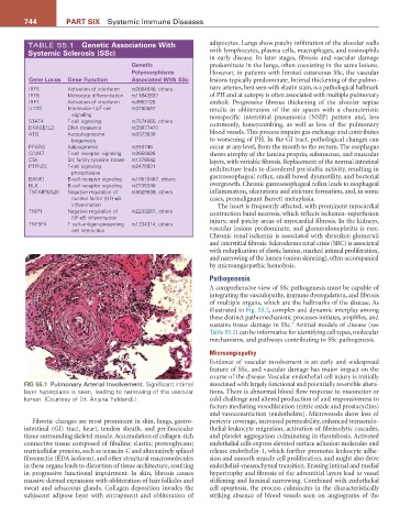
FIG 55.1 Pulmonary Arterial Involvement. Significant intimal associated with largely functional and potentially reversible altera-
layer hyperplasia is seen, leading to narrowing of the vascular tions. There is abnormal blood flow response to vasomotor or
lumen. (Courtesy of Dr. Anjana Yeldandi.) cold challenge and altered production of and responsiveness to
factors mediating vasodilatation (nitric oxide and prostacyclins)
and vasoconstriction (endothelins). Microvessels show loss of
Fibrotic changes are most prominent in skin, lungs, gastro- pericyte coverage, increased permeability, enhanced transendo-
intestinal (GI) tract, heart, tendon sheath, and perifascicular thelial leukocyte migration, activation of fibrinolytic cascades,
tissue surrounding skeletal muscle. Accumulation of collagen-rich and platelet aggregation culminating in thrombosis. Activated
connective tissue composed of fibulins; elastin; proteoglycans; endothelial cells express elevated surface adhesion molecules and
matricellular proteins, such as tenascin-C and alternatively spliced release endothelin-1, which further promotes leukocyte adhe-
fibronectin (EDA isoform), and other structural macromolecules sion and smooth muscle cell proliferation, and might also drive
in these organs leads to distortion of tissue architecture, resulting endothelial–mesenchymal transition. Ensuing intimal and medial
in progressive functional impairment. In skin, fibrosis causes hypertrophy and fibrosis of the adventitial layers lead to vessel
massive dermal expansion with obliteration of hair follicles and stiffening and luminal narrowing. Combined with endothelial
sweat and sebaceous glands. Collagen deposition invades the cell apoptosis, the process culminates in the characteristically
subjacent adipose layer with entrapment and obliteration of striking absence of blood vessels seen on angiograms of the