
Page 846 - Clinical Immunology_ Principles and Practice ( PDFDrive )
P. 846
818 Part SIX Systemic Immune Diseases
A B
C D
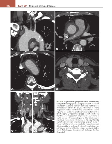
FIG 59.7 Diagnostic Imaging in Takayasu Arteritis (TA):
Computed Tomographic Angiography (CTA). Contrast-
enhanced CTA shows markedly increased wall thickness
of the scanned arteries in a 28-year-old woman. A, Coronal
images demonstrate wall thickening (arrows) of the ascend-
ing aorta, the aortic arch and the proximal aortic branches
(brachiocephalic, left common carotid, and left subclavian).
B and C, Axial images allow for precise measurements of
wall thickness in the ascending aorta (B) and in the aortic
arch (C). D shows marked thickening of the circumferential
wall of both common carotid arteries (arrows). E and F,
Vessel wall inflammation has resulted in moderate stenosis
of both carotid arteries (arrows), with wall irregularities more
pronounces on the right side. [Images were provided by
E F Dr. D. Fleischmann, Department of Radiology, Stanford
University.]