
Page 843 - Clinical Immunology_ Principles and Practice ( PDFDrive )
P. 843
CHaPtEr 59 Large-Vessel Vasculitides 815
$ %
& '
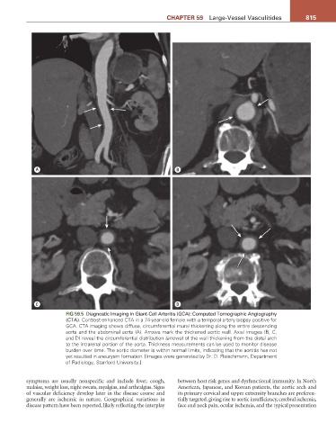
FIG 59.5 Diagnostic Imaging in Giant-Cell Arteritis (GCA): Computed Tomographic Angiography
(CTA). Contrast-enhanced CTA in a 74-year-old female with a temporal artery biopsy positive for
GCA. CTA imaging shows diffuse, circumferential mural thickening along the entire descending
aorta and the abdominal aorta (A). Arrows mark the thickened aortic wall. Axial images (B, C,
and D) reveal the circumferential distribution (arrows) of the wall thickening from the distal arch
to the intrarenal portion of the aorta. Thickness measurements can be used to monitor disease
burden over time. The aortic diameter is within normal limits, indicating that the aortitis has not
yet resulted in aneurysm formation. [Images were generated by Dr. D. Fleischmann, Department
of Radiology, Stanford University.]
symptoms are usually nonspecific and include fever, cough, between host risk genes and dysfunctional immunity. In North
malaise, weight loss, night sweats, myalgias, and arthralgias. Signs American, Japanese, and Korean patients, the aortic arch and
of vascular deficiency develop later in the disease course and its primary cervical and upper extremity branches are preferen-
generally are ischemic in nature. Geographical variations in tially targeted, giving rise to aortic insufficiency, cerebral ischemia,
disease pattern have been reported, likely reflecting the interplay face and neck pain, ocular ischemia, and the typical presentation