
Page 845 - Clinical Immunology_ Principles and Practice ( PDFDrive )
P. 845
CHaPtEr 59 Large-Vessel Vasculitides 817
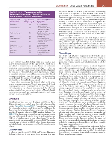
TABLE 59.2 takayasu arteritis: majority of patients. 2,26,34 Generally this is captured by measuring
relationship Between Clinical Symptoms ESR or CRP. It is important to note, however, that a subset of
and affected Vascular territories patients with GCA has normal ESR readings, even before initiation
of immunosuppressive therapy. A normal ESR or CRP reading
Vascular Bed approximate Predominant Clinical is not sufficient to exclude the diagnosis, and further diagnostic
Involvement Frequency (%) Symptoms workup is required if clinical presentation is suspicious for
Subclavian 90 Arm claudication, vasculitis. Other acute-phase proteins, such as fibrinogen and
pulselessness SAA, have been reported to be elevated as well. IL-6 is a potent
Common carotid 60 Visual defects, stroke, inducer of acute-phase proteins in the liver and has been found
transient ischemic to be a sensitive marker of continuous systemic inflammation. 10,11 .
attack, syncope Other laboratory abnormalities, such as elevation of alkaline
Abdominal aorta 45 Claudication, hypertension, phosphatase, thrombocytosis, and anemia, are in line with a
abdominal angina
Renal 35 Hypertension robust acute-phase response.
Aortic arch/root 35 Aortic insufficiency, Autoantibody measurements are not helpful beyond
congestive heart failure excluding differential diagnoses, such as rheumatoid arthritis
Vertebral 35 Dizziness, visual (RA), systemic lupus erythematosus (SLE), or antineutrophil
impairment cytoplasmic antibody (ANCA)–related vasculitides. No disease-
Celiac axis 20 Abdominal angina specific autoantibodies for GCA and TA have been discovered,
Superior mesenteric 20 Abdominal angina emphasizing that B-cell immunity may not contribute to vascular
Iliac 20 Claudication
Pulmonary 10 Dyspnea, chest pain inflammation.
Coronary 10 Myocardial infarction,
angina Tissue Biopsy
In patients with TA, tissue biopsies are rarely available unless
the patient had to undergo vascular reconstructive surgery. In
most patients, the diagnosis is made on the basis of imaging
is now relatively rare, but fleeting visual abnormalities may procedures revealing luminal and wall abnormalities in affected
indicate transient ischemic attacks. Signs of aortic insufficiency blood vessels.
are unlikely to be encountered in early disease, but continuous In contrast, arterial biopsy remains a critical diagnostic
monitoring for aortic dilation is an essential part of follow-up approach in managing patients with GCA. Temporal arteries are
care. Coronary artery stenosis in a young patient must prompt easily accessible, and a segment of these arteries can be removed
the physician to rule out TA. In a subset of patients, the origins in an outpatient setting. Recommendations include harvesting
of mesenteric arteries are involved by stenosing vasculitis. Clinical 2–3 cm of the temporal artery, starting at the most symptomatic
consequences include weight loss, nausea, vomiting, diarrhea, side. Frozen tissue sections can lead to a quick diagnosis of
and abdominal claudication, typically elicited by the increased granulomatous vasculitis. Whether the second side should be
intestinal blood demand following a meal. biopsied during the same surgical procedure remains a matter
Renal artery stenosis may be clinically silent and is often of debate. In cohorts that included several hundred patients,
noticed in routine screening. Correct measurement of blood vasculitis was detected in 2–3% of tissue samples from the second
pressure can represent a pressing clinical problem if the upper- side if the first side was negative. If the clinical suspicion is
extremity arteries are affected. Involvement of the infrarenal strong, biopsy confirmation can be sought from a second-side
aorta can lead to lower-extremity claudication. Musculoskeletal biopsy immediately after the first biopsy or after careful monitor-
examinations are usually unrevealing, although joint and muscle ing of the patient for several weeks. Negative findings on temporal
pains are common complaints. artery biopsy do not exclude the diagnosis of GCA. In a retrospec-
tive cohort study, about half of all patients with subclavian GCA
DIAGNOSIS had no evidence of vasculitis in the temporal arteries, emphasizing
that the disease may display clear preference for certain vascular
35
Classification criteria have been developed for GCA and TA to territories. There has been a recent trend toward considering
differentiate patients with LVV from those with other vasculitic negative biopsies as “false negative” and classifying patients with
entities (Tables 59.3 through 59.5). 31-33 Age at disease onset and a negative biopsy as having GCA. This may lead to unnecessary
the pattern of arteritis are clearly important for establishing the immunosuppressive therapy, and the patient may not thus obtain
diagnosis and distinguishing between these two related vascu- a proper diagnosis. Findings that prompt biopsies (headaches,
lopathies. Diagnostic criteria for PMR remain a challenge (see elevated acute-phase reactants) are notoriously nonspecific, and
Table 59.3) as they rely on nonspecific symptoms, such as muscle both physicians and patients are anxious to avoid treatable
pain and stiffness and elevated erythrocyte sedimentation rate blindness, biasing toward overtreatment. The temporal artery
2
(ESR), all of which can occur in many other diseases. No specific biopsy remains a powerful diagnostic tool and the major tool
laboratory test is currently available to diagnose PMR. Therapeutic that allows unequivocal classification of the disease process. A
responsiveness of patients with PMR to low-dose corticosteroids technically proper temporal artery biopsy will detect vasculitis
is clinically helpful but stresses the need for objective diagnostic in the vast majority of patients.
criteria. Corticosteroid therapy does not eradicate pathological findings
of vascular wall infiltrates, and biopsy can still be valuable in
Laboratory Tests making the diagnosis in patients on steroids. Nevertheless it
21
In all three conditions—GCA, PMR, and TA—the laboratory is possible that treatment with steroids leads to a false-negative
findings indicate an intense acute-phase response in a vast biopsy result in some patients.