
Page 849 - Clinical Immunology_ Principles and Practice ( PDFDrive )
P. 849
CHaPtEr 59 Large-Vessel Vasculitides 821
been disappointing, cautioning one that edema-weighted MR
should not be used as a sole means of measuring disease activity
38
and therapeutic responsiveness. Both, CT angiography and
MRA are now used routinely to monitor progression or regression
of vascular involvement and have an important place in managing
the chronic phase of GCA and TA.
THERAPEUTIC MANAGEMENT
tHEraPEUtIC PrINCIPLES
Treatment of Large Vessel Vasculitides
• To prevent vision loss, patients with giant-cell arteritis (GCA) require
immediate treatment. Similarly, with the threat of catastrophic cerebral
ischemia in Takayasu arteritis (TA), prompt initiation of therapy is
$ imperative.
• Corticosteroids are the immunosuppressive drug of choice for large-
vessel vasculitides (LVVs). Often, the drugs must be given over a
period of several years but may be clinically effective at very low
doses.
• Clinical trials have failed to show convincing steroid-sparing effects
for either methotrexate or tumor necrosis factor (TNF)-α blockade in
GCA.
• A phase 3 clinical trial has demonstrated steroid-sparing effect of
treatment with tocilizumab.
• Molecular studies of the inflammatory infiltrate in GCA have shown
that early and untreated disease is characterized by two functional
T-cell lineages; T helper (Th)1 and Th17 cells. Th17 cells are rapidly
responsive to corticosteroids, whereas Th1 cells persist and promote
chronic, smoldering vasculitis.
• It is not known whether the smoldering activity persisting beyond the
acute phase of the disease requires immunosuppressive therapy or
whether the benefits of chronic immunosuppressive therapy outweigh
the potential risks.
• Clinical experience (not evidence-based therapeutic trials) suggests
that a combination of methotrexate, mycophenolate mofetil, or
TNF-α–blocking agents with corticosteroids may be beneficial in
controlling disease in some patients with TA.
• Close monitoring for diabetes, hypertension, and hyperlipidemia
% combined with bone-saving therapy should be part of the treatment
regime in patients with LVVs on long-term corticosteroids.
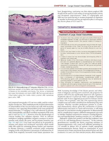
FIG 59.10 Histopathology of Takayasu Arteritis (TA). (A) Full-
thickness section of the aortic wall shows dense mononuclear
infiltrates in the adventitia and media. The intima is thickened With increasing knowledge of the disease process and refine-
and wavy; hematoxylin and eosin (H&E). (B) Florid granulomatous ment of diagnosis and long-term treatment, the prognosis for
inflammation along the media–intima junction with numerous patients with LVVs has significantly improved. Life expectancy
giant cells; H&E. of patients with GCA is preserved. Follow-up studies of Japanese
patients with TA have suggested good control of disease activity
in about 75% of patients, with only 25% experiencing serious
and computed tomography (CT) are now widely used for evaluat- complications and cardiac manifestations dominating long-term
ing the vascular tree. These methods provide excellent information outcome. Whether vasculitis predisposes patients to acceler-
on abnormalities of the vascular lumen and wall, with increasing ated atherosclerotic disease, given the combination of chronic
resolution, now capturing abnormalities in the more peripheral inflammation and injury to vessel wall structures, is still being
arterial branches. CT imaging is fast, well tolerated by patients discussed. It is not known whether progression of atherosclerosis
with claustrophobia, and allows excellent assessment of the aorta and its complications require a different management approach
and its wall (see Fig. 59.7). However, it has the disadvantages of or whether standard vasoprotective measures (treating hyperten-
contrast loading and radiation exposure. With its inherent sion and hyperlipidemia, smoking cessation, etc.) are sufficient. 6
multiplanar imaging capabilities, magnetic resonance is used to Pathogenic studies have pointed out that the traditional view
11
examine neck vessels, the aorta, and its primary branches (see of GCA as a self-limiting disease is incorrect. To the contrary,
Figs. 59.6 and 59.8). Great hope was placed on its potential to granulomatous vasculitis has shown surprising resistance to
measure wall edema and intramural vascularity, which would immunosuppression, with vessel wall infiltrates persisting for
make MR useful for estimating disease burden and responses >12 months in almost 50% of patients despite appropriate
to therapy. However, a carefully conducted study comparing immunosuppressive therapy. Based on examination of serial
imaging results with laboratory parameters of inflammation to temporal artery biopsy specimens from patients before and after
results from surgically harvested vessel biopsy specimens has treatment, it is now clear that arteritis persists, albeit sustained