
Page 844 - Clinical Immunology_ Principles and Practice ( PDFDrive )
P. 844
816 Part SIX Systemic Immune Diseases
A B
C D
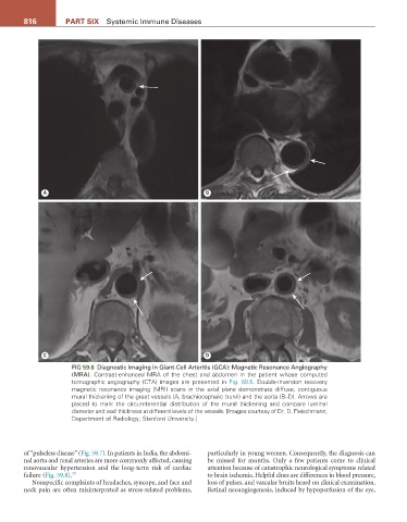
FIG 59.6 Diagnostic Imaging in Giant-Cell Arteritis (GCA): Magnetic Resonance Angiography
(MRA). Contrast-enhanced MRA of the chest and abdomen in the patient whose computed
tomographic angiography (CTA) images are presented in Fig. 59.5. Double-inversion recovery
magnetic resonance imaging (MRI) scans in the axial plane demonstrate diffuse, contiguous
mural thickening of the great vessels (A, brachiocephalic trunk) and the aorta (B–D). Arrows are
placed to mark the circumferential distribution of the mural thickening and compare luminal
diameter and wall thickness at different levels of the vessels. [Images courtesy of Dr. D. Fleischmann,
Department of Radiology, Stanford University.]
of “pulseless disease” (Fig. 59.7). In patients in India, the abdomi- particularly in young women. Consequently, the diagnosis can
nal aorta and renal arteries are more commonly affected, causing be missed for months. Only a few patients come to clinical
renovascular hypertension and the long-term risk of cardiac attention because of catastrophic neurological symptoms related
failure (Fig. 59.8). 30 to brain ischemia. Helpful clues are differences in blood pressure,
Nonspecific complaints of headaches, syncope, and face and loss of pulses, and vascular bruits heard on clinical examination.
neck pain are often misinterpreted as stress-related problems, Retinal neoangiogenesis, induced by hypoperfusion of the eye,