
Page 89 - Clinical Immunology_ Principles and Practice ( PDFDrive )
P. 89
74 Part one Principles of Immune Response
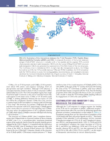
T cell
TCRα
TCRβ
CD4
MHC
Class I
-m
β 2
CD8
Antigen-presenting cell
FIG 4.15 Illustration of the Interactions between the T-Cell Receptor (TCR), Peptide Major
Histocompatibility Complex (pMHC), and CD8. A composite illustration of the human leukocyte
antigen (HLA)-A*0201 structure in complex with a Tax peptide and its cognate TCR α and β
chains (protein data bank (pdb) designation 1BD2) with the human CD8αα/HLA-A*0201 structure
(pdb designation 1AKJ) was generated by superposition of the HLA moiety of the two structures.
The HLA heavy chain is indicated as MHC, its light chain (β 2 microglobulin) as β 2 -m, the CD8αα
homodimer as CD8, the TCR α and β chains as TCRα and TCRβ. In addition, the CD4 homodimer
(pdb file 1WIO) is shown to scale. Connecting peptides, transmembrane, and cytoplasmic domains
are drawn by hand and indicated by dotted lines. (Figure courtesy of David H. Margulies, National
Institute of Allergy and Infectious Diseases, National Institutes of Health.)
CD8α, a 34- to 37-kDa protein, and CD8β, a 32-kDa protein, similar to that of the crystal structure of TCRαβ–pMHC–CD4,
share about 20% amino acid sequence homology. Both are with pMHC at the apex of the “V” and the TCR and CD8 forming
glycoproteins and IgSF members. Although CD8 subserves a the arms of the “V.” CD8 binds to pMHC with lower affinity
coreceptor function similar to that of CD4, in structure it differs and with faster kinetics compared with the TCR. Thus the binding
substantially from CD4. The CD8 extracellular regions have single properties of the CD8 coreceptor, like those of CD4, are consistent
N-terminal IgSF V domains at the end of extended mucin-like with a model in which the TCR initiates pMHC binding, followed
stalk regions of 48 amino acids (CD8α) or 35–38 amino acids by engagement of CD8 to the same pMHC. 66,69-72
(CD8β). A striking difference between the two forms of CD8
lies within the cytoplasmic domain. CD8α, like CD4, contains COSTIMULATORY AND INHIBITORY T-CELL
a cysteine-based motif that enables it to interact with LCK through MOLECULES: THE CD28 FAMILY
a “zinc clasp”–like structure. In contrast, CD8β lacks this motif
and does not associate with LCK. Interestingly, CD8αβ appears Although the T-cell response to antigen requires the binding
to be a more effective activator of TCR signaling than CD8αα. of the TCR and its coreceptors to pMHC, additional receptor–
This may reflect the palmitoylation of the cytoplasmic domain ligand interactions affect the outcome by delivering signals that
of CD8β, which allows CD8αβ to associate with lipid rafts during promote activation (costimulation) or that inhibit it (Table 4.4).
T-cell activation. 66,69,71,72 Prominent among these are the interactions of members of the
73
The structure of CD8αα–pMHC class I complexes demon- CD28 family with their cell-surface ligands on APCs. This family
strates that CD8αα binds to conserved residues in the α 3 domain includes CD28, inducible T-cell costimulator (ICOS), cytotoxic
of MHC class I (i.e., a nonpolymorphic, membrane-proximal T lymphocyte antigen-4 (CTLA-4), B- and T lymphocyte attenu-
region of the molecule distinct from the peptide-binding groove ator (BTLA), and programmed death 1 (PD-1). CD28 and ICOS
engaged by the TCR) (Chapter 5). Compared with the interaction are costimulatory receptors; the major functions of CTLA-4,
of CD4 and MHC class II, binding is more antibody-like, with PD-1, and BTLA are inhibitory. CD28 and CTLA-4 are T-cell
a loop of the MHC α 3 domain locked between the CDR-like specific, whereas BTLA and PD-1 are also expressed by B cells
loops of the two CD8α IgSF V domains. Models of the structure and ICOS by NK cells. CD28, CTLA-4, and PD-1 are the targets
of the TCRαβ–pMHC–CD8 ternary complex propose a “V” shape of therapeutic interventions in current clinical practice.