
Page 87 - Clinical Immunology_ Principles and Practice ( PDFDrive )
P. 87
72 Part one Principles of Immune Response
A Lck binding site
ITAM
D1 CD4 TCR CD8
D2 α β
CD3 CD3
D3 α β
ε δ γ ε
D4 ς ς
- - + + + - - - -
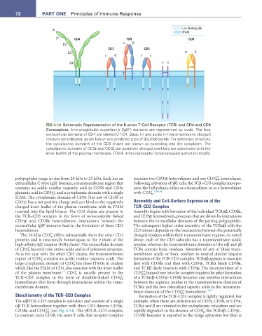
FIG 4.14 Schematic Representation of the Human T-Cell Receptor (TCR) and CD4 and CD8
Coreceptors. Immunoglobulin superfamily (IgSF) domains are represented by ovals. The four
extracellular domains of CD4 are labeled D1-D4. Basic (+) and acidic (−) transmembrane charged
residues are indicated, as are known and predicted sites of disulfide bonds. For schematic simplicity
the cytoplasmic domains of the CD3 chains are shown as extending into the cytoplasm. The
cytoplasmic domains of CD3ε and CD3ζ are positively charged and likely are associated with the
inner leaflet of the plasma membrane. ITAM, immunoreceptor tyrosine-based activation motifs.
polypeptides range in size from 20 kDa to 25 kDa. Each has an contains two CD3γε heterodimers and one CD3ζζ homodimer.
extracellular C-type IgSF domain, a transmembrane region that Following activation of γδT cells, the TCR–CD3 complex incorpo-
contains an acidic residue (aspartic acid in CD3δ and CD3ε rates the FcRγ chain, either as a homodimer or as a heterodimer
glutamic acid in CD3γ), and a cytoplasmic domain with a single with CD3ζ. 18,60,61
ITAM. The cytoplasmic domain of CD3ε (but not of CD3δ or
CD3γ) has a net positive charge and can bind to the negatively Assembly and Cell-Surface Expression of the
charged inner leaflet of the plasma membrane with its ITAM TCR–CD3 Complex
inserted into the lipid bilayer. The CD3 chains are present in Assembly begins with formation of the individual TCRαβ, CD3δε,
the TCR–CD3 complex in the form of noncovalently linked and CD3γε heterodimers, processes that are driven by interactions
CD3γε and CD3δε heterodimers; interactions between the between the extracellular domains of the pairing polypeptides.
extracellular IgSF domains lead to the formation of these CD3 The subsequent higher order assembly of the TCRαβ with the
heterodimers. CD3 dimers depends on the interactions between the potentially
The 16 kDa CD3ζ differs substantially from the other CD3 charged residues within their transmembrane regions. As noted
proteins and is structurally homologous to the γ chain of the above, each of the CD3 subunits has a transmembrane acidic
high-affinity IgE receptor (FcRγ chain). The extracellular domain residue, whereas the transmembrane domains of the αβ and γδ
of CD3ζ has only nine amino acids and is of unknown structure. TCRs contain basic residues. Mutation of any of these trans-
As is the case with the other CD3 chains, the transmembrane membrane acidic or basic residues to neutral alanine impairs
region of CD3ζ contains an acidic residue (aspartic acid). The formation of the TCR–CD3 complex. TCRαβ appears to associate
large cytoplasmic domain of CD3ζ has three ITAMs in tandem first with CD3δε and then with CD3γε. TCRα binds CD3δε,
which, like the ITAM of CD3, also associate with the inner leaflet and TCRβ likely interacts with CD3γε. The incorporation of a
18
of the plasma membrane. CD3ζ is usually present in the CD3ζζ homodimer into the complex requires the prior formation
TCR–CD3 complex in the form of disulfide-linked CD3ζζ of a TCRαβ–CD3γε–CD3δε hexamer and involves interactions
homodimers that form through interactions within the trans- between the arginine residue in the transmembrane domain of
membrane domain. TCRα and the two colocalized aspartic acids in the transmem-
brane domains of the CD3ζζ homodimer. 18,62
Stoichiometry of the TCR–CD3 Complex Formation of the TCR–CD3 complex is tightly regulated. For
The αβTCR–CD3 complex is univalent and consists of a single example, when there are deficiencies of CD3γ, CD3δ, or CD3ε,
αβ TCR heterodimer together with three CD3 dimers: CD3γε, TCRα and β are retained in the endoplasmic reticulum and are
CD3δε, and CD3ζζ (see Fig. 4.14). The γδTCR–CD3 complex, rapidly degraded. In the absence of CD3ζ, the TCRαβ–CD3γε–
in contrast, lacks CD3δ. On naïve T cells, this receptor complex CD3δε hexamer is exported to the Golgi apparatus but then is