
Page 957 - Clinical Immunology_ Principles and Practice ( PDFDrive )
P. 957
924 Part Seven Organ-Specific Inflammatory Disease
$ &
% '
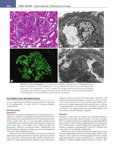
FIG 68.9 C3 Glomerulopathy. A, Glomerulus exhibiting the characteristic membranoproliferative
pattern (hematoxylin and eosin [H&E] stain). B, Immunofluorescence showing prominent glomerular
staining for C3 complement. C and D, Electron microscopy ultrastructure showing mesangial
interposition giving a double contour of the glomerular capillary wall, as well as interposed electron
dense deposits in the subendothelial space and the mesangium.
POSTINFECTIOUS NEPHROPATHIES entacavir, adefovir, tenofovir, and telbivudine. Treatment with
nucleoside/nucleotide analogues are generally used for at least
An ever-expanding list of infectious agents has been implicated 1 year and continued for at least 6 months after HBeAg sero-
in the pathogenesis of certain forms of immune-mediated conversion. Immunosuppression (corticosteroids with or without
nephropathies. cyclophosphamide or rituximab) may cautiously be considered
in patients with rapidly progressive glomerulonephritis, but
Viral Infections concomitant antiviral therapy is required.
Hepatitis B
Hepatitis B–associated nephropathy most frequently presents as Hepatitis C
nephrotic syndrome accompanied by microscopic hematuria; Hepatitis C virus (HCV) is a major cause of both transfusion-
renal biopsy most commonly shows MN. Immunofluorescence associated and sporadic chronic hepatitis. The most common
studies usually show Ig, C3, and some IgM staining in the HCV-associated renal disease is usually, but not invariably, MPGN
subepithelial region along the glomerular basement membranes in the context of type II mixed cryoglobulinemia. The pathogenesis
and in some cases viral antigens can be detected within the of the renal disease is caused by deposition within glomeruli of
glomerulus. Electron microscopy shows subepithelial and immune complexes containing HCV, anti-HCV antibody and
intramembranous deposits, but there may also be mesangial and virus-related (or unrelated) cryoglobulins.
even some subendothelial deposits. Treatment of HCV infection has greatly improved in the last
Therapy for hepatitis B renal disease has focused on antiviral few years. Historically, treatment was mainly based on IFN-α and
drugs because glucocorticoids and cytotoxic agents may promote ribavirin. These agents are now being replaced by HCV-specific
viral replication. Treatment generally consists of interferon-α antiviral drugs that are used in combination (i.e., sofosbuvir plus
23
(IFN-α) or nucleoside/nucleotide analogues, such as lamividine, ledipasvir), which are highly effective and often curative. The