
Page 96 - Clinical Immunology_ Principles and Practice ( PDFDrive )
P. 96
CHAPtER 5 The Major Histocompatibility Complex 81
KEY CoNCEPtS Peptide
Genomic Organization of the MHC
α 1 α 2 α 1 β 1
• The major histocompatibility complex (MHC) is the most complex
genomic region in the whole human genome. It is associated with
more diseases than any other genomic region of comparable size.
• The class I region contains the polymorphic human leukocyte antigen β m α 3 α 2 β 2
2
(HLA)-A, -B, and -C genes; the less polymorphic nonclassic class I
HLA-E, HLA-F, and HLA-G genes; and the class I-related MICA and
MICB.
• The class II region contains the HLA-DR A and B, DQ A and B, and Cell membrane
DP A and B genes. It also contains the TAP, LMP, DM, and DO genes,
which encode molecules that help process antigens into peptides
that can bind class I and class II molecules.
• Genes within the MHC demonstrate extensive linkage disequilibrium. HLA class I molecule HLA class II molecule
A string of alleles of polymorphic MHC genes that commonly exist
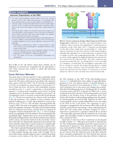
in linkage disequilibrium within a given population is termed an MHC FIG 5.2 Human Leukocyte Antigen (HLA) Class I and II Domain
haplotype. Organization. Although HLA class I and class II proteins have
• Haplotypes are preserved by means of natural selection, which a different chain structure, the organization of their domains is
acts to secure survival advantages for reproductive fitness within extremely similar. Both class I and II molecules are expressed
a given environment. on the cell surface, where they are accessible to T cells. Both
• Common haplotypes within a given population appears to reflect
functional interdependencies of the MHC gene alleles. have an outermost domain that contains a cleft where antigenic
• Haplotypes can be different in different populations. peptides are displayed. Two of the three class I α domains fold
• The HLA genes of the two chromosomes (haplotypes) are to create a domain with a peptide-binding cleft. The remaining
coexpressed. α 3 domain helps support the peptide binding domain and anchors
the molecular to the cell membrane. The class I molecule also
contains an extrinsic β chain, β 2 microglobulin, which is encoded
by a separate, invariant gene. β 2 microglobulin associates with
then traffic to the cell surface, where these peptides can be the α 3 domain to support the antigen-binding domain created
displayed, or presented, for recognition by the appropriate T by the α 1 and α 2 domains. Class II molecules share a similar
cells. Their structure has evolved to satisfy this particular overall structure but are the product of two genes of variable
requirement. sequence, each of which contains one α and one β domain.
Classic HLA Class I Molecules
The classic HLA-A, HLA-B, and HLA-C class I molecules consist
of an α and a β chains. The α chain masses 45 kilodalton (kDa) the NH 2 terminus to the “left” of the HLA-binding groove
and is 362-366 amino acids long. It is encoded by the respective (see Fig. 5.3). Individual HLA class I alleles are generally distin-
class I genes within the MHC. The β chain, β 2 microglobulin guished by their own distinct pattern of peptide binding, as
5
(12 kDa), is encoded by its respective gene on chromosome 15. illustrated for selected HLA-B molecules in Table 5.1. Among
The α chain has three ≈90 amino acid extracellular domains class I molecules, one or a few amino acid changes may consider-
encoded by exons 2, 3, and 4, respectively, a transmembrane ably alter the binding properties of a binding pocket. In a healthy
segment (≈ 25 amino acids) encoded by exon 5 and a C-terminal nonendocytosing cell, HLA molecules are filled with a variety
cytoplasmic end (≈30 amino acids) encoded by exons 6 and 7. β 2 of peptides from self molecules. The bound peptides are selected
microglobulin, which is invariant, comprises the fourth domain according to the binding motif of the particular allele. Even
(Fig. 5.2). The first two α domains (α 1 and α 2 ) are the most distal during viral infection or upon pathogen phagocytosis, the number
to the cell membrane. They combine to form a domain with a of nonself peptides may not be high. Together, the MHC class
peptide-binding groove, or cleft, that is flanked by a surface that I and its peptide create a complex ligand that serves as the target
interacts with a TCR or a NK cell killer immunoglobulin-like of the TCR on the T-cell surface. The expression of class I
receptor (KIR). The ends of the peptide-binding cleft are closed molecules is upregulated by the interferons (IFNs) IFN-α, IFN-β,
and fix the peptide’s orientation. The sides of the peptide-binding and IFN-γ, granulocyte macrophage–colony-stimulating factor
cleft are composed of α helices, and the floor is composed of (GM-CSF) and certain other cytokines (Chapter 9). Class I
symmetric strands of β pleated sheet (Fig. 5.3). The α 3 domain expression is governed by a regulatory element that is located
and β 2 microglobulin are both members of the immunoglobulin ≈160 nucleotides upstream from the initiation site of the class
superfamily (IgSF). Together they create a structure that supports I gene. This site binds a number of regulatory factors, including
the peptide-binding domain and, with the transmembrane domain those induced by IFNs.
of the α chain, attaches the molecule to the cell surface. Class I Intact HLA-A, HLA-B, or HLA-C molecules are also ligands
6,7
HLA molecules are ubiquitously expressed in all nucleated cells for KIR (Chapter 17). KIR genes are located on chromosome
and in platelets. Expression of class I molecules is significantly 19. Their independent segregation from HLA genes located on
reduced on red blood cells (RBCs) and absent on sperm cells. chromosome 6 produces a wide diversity in the number and
HLA class I molecules bind peptides derived from the processed type of inherited HLA–KIR combinations. These combinations
proteins of a pathogen or other self/nonself peptides (Chapter eventually influence immune competency and adaptability. KIRs
6). These peptides average nine amino acids in length. Two or do not interact with the whole top area of the HLA molecule
more of the amino acid side chains are used to anchor the peptide that is normally recognized by the TCR. Instead, they interact
to pockets on the surface of the HLA class I molecule, with with one end of the top of the molecule. All HLA-C alleles are