
Page 1168 - Hall et al (2015) Principles of Critical Care-McGraw-Hill
P. 1168
CHAPTER 86: Intracranial Pressure: Monitoring and Management 807
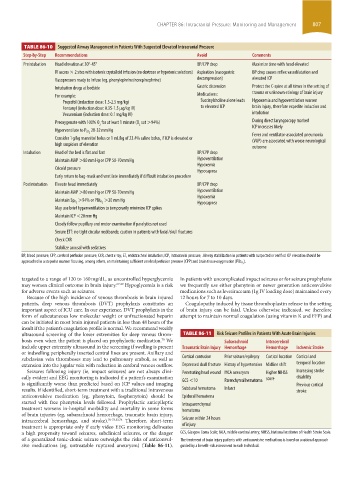
TABLE 86-10 Suggested Airway Management in Patients With Suspected Elevated Intracranial Pressure
Step-by-Step Recommendations Avoid Comments
Preintubation Head elevation at 30°-45° BP/CPP drop Maximize time with head elevated
IV access × 2 sites with isotonic crystalloid infusion (no dextrose or hypotonic solutions) Aspiration (nasogastric BP drop causes reflex vasodilatation and
Vasopressors ready to infuse (eg, phenylephrine/norepinephrine) decompression) elevated ICP
Intubation drugs at bedside Gastric distension Protect the C-spine at all times in the setting of
For example: Medications: trauma or unknown etiology of brain injury
Propofol (induction dose: 1.5-2.5 mg/kg) Succinylcholine alone leads Hypoxemia and hypoventilation worsen
Fentanyl (induction dose: 0.35-1.5 µg/kg IV) to elevated ICP brain injury, therefore expedite induction and
Vecuronium (induction dose: 0.1 mg/kg IV) intubation
Preoxygenate with 100% O for at least 1 minute (O sat >94%) During direct laryngoscopy marked
2 2 ICP increases likely
28-32 mm Hg
Hyperventilate to P CO 2
Fever and ventilator-associated pneumonia
Consider 1 g/kg mannitol bolus or 1 mL/kg of 23.4% saline bolus, if ICP is elevated or (VAP) are associated with worse neurological
high suspicion of elevation
outcome
Intubation Head of the bed is flat and fast BP/CPP drop
Maintain MAP >80 mm Hg or CPP 50-70 mm Hg Hypoventilation
Hypoxemia
Cricoid pressure Hypocapnea
Early return to bag-mask and ventilate immediately if difficult intubation procedure
Postintubation Elevate head immediately BP/CPP drop
Maintain MAP >80 mm Hg or CPP 50-70 mm Hg Hypoventilation
Hypoxemia
>20 mm Hg
Hypocapnea
Maintain Sp O 2 >94% or PBt O 2
May use brief hyperventilation to temporarily minimize ICP spikes
Maintain ICP <20 mm Hg
Closely follow pupillary and motor examination if paralytics not used
Secure ETT: no tight circular neckbands; caution in patients with facial/skull fractures
Check CXR
Stabilize arousal with sedatives
BP, blood pressure; CPP, cerebral perfusion pressure; CXR, chest x-ray; ET, endotracheal intubation; ICP, intracranial pressure; .Airway stabilization in patients with suspected or verified ICP elevation should be
approached in a stepwise manner focusing, among others, on maintaining sufficient cerebral perfusion pressure (CPP) and brain tissue oxygenation (PBtO ).
2
targeted to a range of 120 to 160 mg/dL, as uncontrolled hyperglycemia In patients with uncomplicated impact seizures or for seizure prophylaxis
may worsen clinical outcome in brain injury. 67-69 Hypoglycemia is a risk we frequently use either phenytoin or newer generation anticonvulsive
for adverse events such as seizures. medications such as levetiracetam (1g IV loading dose) maintained every
Because of the high incidence of venous thrombosis in brain injured 12 hours for 7 to 10 days.
patients, deep venous thrombosis (DVT) prophylaxis constitutes an Coagulopathy induced by tissue thromboplastin release in the setting
important aspect of ICU care. In our experience, DVT prophylaxis in the of brain injury can be fatal. Unless otherwise indicated, we therefore
form of subcutaneous low molecular weight or unfractionated heparin attempt to maintain normal coagulation (using vitamin K and FFP) and
can be initiated in most brain injured patients in less than 48 hours of the
insult if the patient’s coagulation profile is normal. We recommend weekly
ultrasound screening of the lower extremities for deep venous throm- TABLE 86-11 Risk Seizure Profiles in Patients With Acute Brain Injuries
bosis even when the patient is placed on prophylactic medication. We Subarachnoid Intracerebral
70
include upper extremity ultrasound in the screening if swelling is present Traumatic Brain Injury Hemorrhage Hemorrhage Ischemic Stroke
or indwelling peripherally inserted central lines are present. Axillary and
subclavian vein thromboses may lead to pulmonary emboli, as well as Cortical contusion Prior seizure/epilepsy Cortical location Cortical and
extension into the jugular vein with reduction in cerebral venous outflow. Depressed skull fracture History of hypertension Midline shift temporal location
Seizures following injury (ie, impact seizures) are not always clini- Penetrating head wound MCA aneurysm Higher NIHSS Increasing stroke
cally evident and EEG monitoring is indicated if a patient’s examination score disability
is significantly worse than predicted based on ICP values and imaging GCS <10 Parenchymal hematoma Previous cortical
results. If identified, short-term treatment with a traditional intravenous Subdural hematoma Infarct stroke
anticonvulsive medication (eg, phenytoin, fosphenytoin) should be Epidural hematoma
started with free phenytoin levels followed. Prophylactic antiepileptic Intraparenchymal
treatment worsens in-hospital morbidity and mortality in some forms hematoma
of brain injuries (eg, subarachnoid hemorrhage, traumatic brain injury,
intracerebral hemorrhage, and stroke). 70-73,33,74 Therefore, short-term Seizure within 24 hours
treatment is appropriate only if early video EEG monitoring delineates of injury
a high propensity toward seizures, subclinical seizures, or the danger GCS, Glasgow Coma Scale; MCA, middle cerebral artery; NIHSS, National Institutes of Health Stroke Scale.
of a generalized tonic-clonic seizure outweighs the risks of anticonvul- The treatment of brain injury patients with anticonvulsive medications is based on a rational approach
sive medications (eg, untreatable ruptured aneurysm) (Table 86-11). guided by a benefit-risk assessment in each individual.
section06.indd 807 1/23/2015 12:56:06 PM