
Page 1546 - Hall et al (2015) Principles of Critical Care-McGraw-Hill
P. 1546
CHAPTER 112: Principles of Postoperative Critical Care 1065
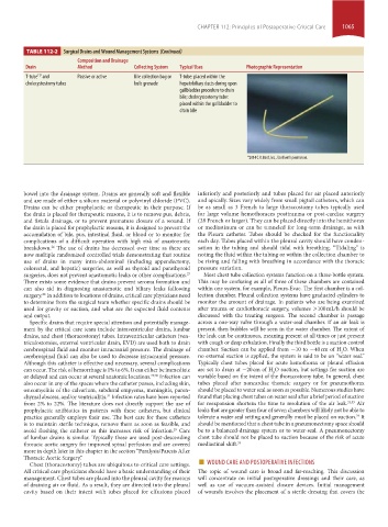
TABLE 112-2 Surgical Drains and Wound Management Systems (Continued)
Composition and Drainage
Drain Method Collecting System Typical Uses Photographic Representation
T-tube and Passive or active Bile collection bag or T-tube: placed within the
117
cholecystostomy tubes bulb grenade hepatobiliary ducts during open
gallbladder procedure to drain
bile; cholecystostomy tube:
placed within the gallbladder to
drain bile
®2014 C.R. Bard, Inc., Used with permission.
bowel into the drainage system. Drains are generally soft and flexible inferiorly and posteriorly and tubes placed for air placed anteriorly
and are made of either a silicon material or polyvinyl chloride (PVC). and apically. Sizes vary widely from small pigtail catheters, which can
Drains can be either prophylactic or therapeutic in their purpose. If be as small as 3 French to large thoracostomy tubes typically used
the drain is placed for therapeutic reasons, it is to remove pus, debris, for large volume hemothoraces posttrauma or post-cardiac surgery
and fistula drainage, or to prevent premature closure of a wound. If (28 French or larger). They can be placed directly into the hemithorax
the drain is placed for prophylactic reasons, it is designed to prevent the or mediastinum or can be tunneled for long-term drainage, as with
accumulation of bile, pus, intestinal fluid, or blood or to monitor for the Pleurx catheter. Tubes should be checked for the functionality
complications of a difficult operation with high risk of anastomotic each day. Tubes placed within the pleural cavity should have conden-
breakdown. The use of drains has decreased over time as there are sation in the tubing and should tidal with breathing. “Tidaling” is
26
now multiple randomized controlled trials demonstrating that routine noting the fluid within the tubing or within the collection chamber to
use of drains in many intra-abdominal (including appendectomy, be rising and falling with breathing in accordance with the thoracic
colorectal, and hepatic) surgeries, as well as thyroid and parathyroid pressure variation.
surgeries, does not prevent anastomotic leaks or other complications. Most chest tube collection systems function on a three-bottle system.
27
There exists some evidence that drains prevent seroma formation and This may be confusing as all of three of these chambers are contained
can also aid in diagnosing anastomotic and biliary leaks following within one system, for example, Pleura-Evac. The first chamber is a col-
surgery. In addition to locations of drains, critical care physicians need lection chamber. Pleural collection systems have graduated cylinders to
28
to determine from the surgical team whether specific drains should be monitor the amount of drainage. In patients who are being examined
used for gravity or suction, and what are the expected fluid contents after trauma or cardiothoracic surgery, volumes >100 mL/h should be
and output. discussed with the treating surgeon. The second chamber is passage
Specific drains that require special attention and potentially manage- across a one-way valve through a water-seal chamber. If an air leak is
ment by the critical care team include intraventricular drains, lumbar present, then bubbles will be seen in the water chamber. The extent of
drains, and chest (thoracostomy) tubes. Intraventricular catheters (ven- the leak can be continuous, meaning present at all times or just present
triculostomies, external ventricular drain, EVD) are used both to drain with cough or deep exhalation. Finally the third bottle is a suction control
cerebrospinal fluid and monitor intracranial pressure. The drainage of chamber. Suction can be applied from −10 to −40 cm of H O. When
2
cerebrospinal fluid can also be used to decrease intracranial pressure. no external suction is applied, the system is said to be on “water seal.”
Although this catheter is effective and necessary, several complications Typically chest tubes placed for acute hemothorax or pleural effusion
can occur. The risk of hemorrhage is 1% to 6%. It can either be immediate are set to drain at −20 cm of H O suction, but settings for suction are
2
or delayed and can occur at several anatomic locations. 29,30 Infection can variable based on the intent of the thoracostomy tube. In general, chest
also occur in any of the spaces where the catheter passes, including skin, tubes placed after noncardiac thoracic surgery or for pneumothorax
osteomyelitis of the calvarium, subdural empyema, meningitis, paren- should be placed to water seal as soon as possible. Numerous studies have
chymal abscess, and/or ventriculitis. Infection rates have been reported found that placing chest tubes on water seal after a brief period of suction
31
from 2% to 22%. The literature does not directly support the use of for reexpansion shortens the time to resolution of the air leak. 32,33 Air
prophylactic antibiotics in patients with these catheters, but clinical leaks that are greater than four of seven chambers will likely not be able to
practice generally employs their use. The best care for these catheters tolerate a water seal setting and generally must be placed on suction. It
34
is to maintain sterile technique, remove them as soon as feasible, and should be mentioned that a chest tube in a pneumonectomy space should
avoid flushing the catheter as this increases risk of infection. Care be to a balanced-drainage system or to water-seal. A pneumonectomy
29
of lumbar drains is similar. Typically these are used post-descending chest tube should not be placed to suction because of the risk of acute
thoracic aortic surgery for improved spinal perfusion and are covered mediastinal shift. 35
more in depth later in this chapter in the section “Paralysis/Paresis After
Chest (thoracostomy) tubes are ubiquitous to critical care settings. ■
Thoracic Aortic Surgery.” WOUND CARE AND POSTOPERATIVE INFECTIONS
All critical care physicians should have a basic understanding of their The topic of wound care is broad and far-reaching. This discussion
management. Chest tubes are placed into the pleural cavity for reasons will concentrate on initial postoperative dressings and their care, as
of draining air or fluid. As a result, they are directed into the pleural well as use of vacuum-assisted closure devices. Initial management
cavity based on their intent with tubes placed for effusions placed of wounds involves the placement of a sterile dressing that covers the
section10.indd 1065 1/20/2015 9:19:41 AM