
Page 1606 - Hall et al (2015) Principles of Critical Care-McGraw-Hill
P. 1606
CHAPTER 118: Head Injury 1125
Response Scale RESPIRATORY MANAGEMENT
Eye opening Hypoxemia and hypotension are the two most important factors associ-
ated with adverse outcomes in patients after TBI, and the association
None 1
with TBI is stronger than in trauma patients without neurological
To pain 2 injury. 15,16 Patients who have severe brain injury are at increased risk for
17
To voice 3 acute respiratory distress syndrome (ARDS). Patients with severe head
injury (GCS ≤8) may have an abnormal lung elasticity and resistance
Spontaneous 4 as early as day 1 post injury. A recent retrospective cohort study of the
18
Best verbal response Nationwide Inpatient Sample (NIS) database reported a 22% prevalence
of ARDS/acute lung injury (ALI) after TBI in 2008 with an in-hospital
None 1
ARDS/ALI-related mortality of 28%. 19
Incomprehensible 2 Hypoxemia may be caused by noncardiogenic pulmonary edema
Inappropriate 3 from ARDS due to a systemic inflammatory response to trauma or fat
emboli, neurogenic pulmonary edema, or less commonly, cardiogenic
Confused 4 pulmonary edema. Other etiologies of hypoxemia include airway
Oriented, normal conversation 5 obstruction, lung contusion from direct chest trauma, flail chest,
pneumothorax, retained secretions or aspiration, pneumonia, and
Best motor response
hypercarbia. Hypercarbia may be caused by depressed respirations from
None 1 coma or brain stem dysfunction, chest trauma, airway obstruction, or
high cervical spine injuries.
Extension to pain (decerebrate) 2
Oxygenation should be monitored by pulse oximetry and checked by
Flexion to pain (decorticate) 3 <60 mm Hg or hemoglo-
arterial blood gases. Hypoxemia defined as Pa O 2
15
Withdrawal to pain 4 bin-oxygen saturation <90% must be avoided. After TBI, patients with
any of the following: signs of respiratory distress, intracranial hyperten-
Localizes pain 5 sion, impending herniation, encephalopathy or coma (GCS ≤9), requiring
Obeys commands 6 high levels of inspired oxygen to maintain Pa O 2 above 60 mm Hg, absolute
CO retention, or CO retention relative to respiratory minute volume
2
2
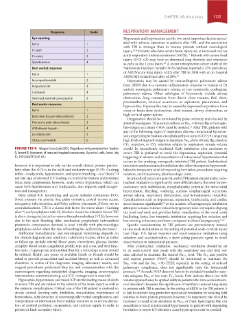
FIGURE 118-9. Glasgow Coma Scale (GCS). (Reproduced with permission from Teasdale should be immediately intubated. Early intubation after moderate to
G, Jennett B. Assessment of coma and impaired consciousness. A practical scale, Lancet. July severe TBI is preferred to avoid the hypoxemia, aspiration, potential
13, 1974;304(7872):81-84.) triggering of seizures and exacerbation of intracranial hypertension that
occurs in the crashing, emergently intubated TBI patient. Endotracheal
however, it is important to rely on the overall clinical picture, particu- intubation and mechanical ventilation also allow therapeutic hyperventi-
larly when the GCS is in the mild and moderate range (9-15). Cushing lation for temporary relief of impending herniation, procedures requiring
reflex—bradycardia, hypertension, and apneic breathing—is a “classic” if sedation, and if necessary, pharmacologic coma.
not late sign of elevated ICP leading to cerebral herniation and terminal In critically ill patients in general, and in TBI patients in particular, endo-
brain stem compression; however, acute severe hypoxemia, which can tracheal intubation is significantly more difficult due to the need for pre-
cause both hypertension and bradycardia, also requires rapid recogni- cautionary neck stabilization, encephalopathy, potential for intracranial
tion and management. hypertension, bleeding, vomiting, copious oropharyngeal secretions,
Basic initial ICU monitoring and access includes continuous ECG, airway edema, respiratory dysfunction, and hemodynamic instability.
blood pressure via arterial line, pulse oximetry, central venous access, Complications such as hypoxemia, aspiration, bradycardia, and cardiac
nasogastric tube insertion, and Foley catheter placement, if there are no arrest increase significantly as the number of laryngoscopic intubation
20
contraindications. TBI is a classic risk factor for stress ulcers (Cushing attempts increase. Indirect optical laryngoscopy does not require aligning
ulcer ) and prophylaxis with H -blockers should be initiated. Severe TBI the head and neck and provides better visualization of the vocal cords
14
2
is also a strong risk factor for venous thromboembolism (VTE); however, facilitating faster, less traumatic intubation requiring less sedation and
due to the early bleeding risks, mechanical prophylaxis (intermittent less training to become proficient compared to direct laryngoscopy. 21,22
pneumatic compression devices) is used initially with pharmacologic Specific considerations in TBI patients are precautionary manual
prophylaxis added when the risk of bleeding has sufficiently decreased. in-line neck stabilization in the setting of potential acute cervical injury
Additional hemodynamic and neurological monitoring depends on (see Chap. 119, Spinal Injuries) and rapid sequence intubation using
the clinical diagnosis and condition. Laboratory studies, either as initial sedatives and succinylcholine, a short acting paralytic agent, to avoid
or follow-up, include arterial blood gases, electrolytes, glucose, lactate, exacerbations in intracranial pressure.
complete blood count, coagulation profile, type and cross, and liver func- After endotracheal intubation, mechanical ventilation should be set
tion tests. If appropriate and omitted thus far, a toxicology screen should to an assist-control type mode with the respiratory rate and tidal vol-
be ordered. Health care proxy or available family or friends should be ume adjusted to maintain the desired Pa CO 2 level. The Fi O 2 and positive
asked to provide preaccident and accident history as well as advanced end expired pressure (PEEP) should be minimized to maintain the
directives. A review of the diagnostic imaging, laboratory results, and Pa O 2 >60 and the Sa O 2 >90. PEEP, especially in the setting of reduced
surgical procedures performed thus far, and communication with the pulmonary compliance, does not significantly raise the intracranial
neurosurgeon regarding anticipated diagnostic imaging, neurosurgical pressure. 23,24 As such, PEEP does not have to be avoided if needed to main-
interventions, neuromonitoring, and ICU management is essential. tain adequate Pa O 2 at less toxic Fi O 2 levels. Data indicate that a low tidal
Hypoxemia, hypotension, and raised ICP are the leading causes of death volume approach may be applied safely in patients who have acute intracra-
in severe TBI and are related to the severity of the brain injury as well as nial disorders ; however, the significance of ventilator-induced lung injury
25
the systemic complications. Critical care of the TBI patient is centered on in patients with TBI is unclear. In the setting of ARDS in the TBI patient, it
airway control, favoring early intubation, resuscitation, maintenance of is safe to institute lung-protective mechanical ventilation by reducing tidal
homeostasis, early detection of neurosurgically treated complications, and volumes to lower plateau pressures; however, the respiratory rate should be
interpretation of information from bedside monitors to minimize disrup- increased to avoid acute elevations in Pa CO 2 or frank hypercapnia that can
tion of cerebral perfusion, oxygenation, and nutrient supply in order to exacerbate or result in intracranial hypertension. In patients with impending
prevent or limit secondary injury. herniation or severe ICP elevation, acute hypercapnia must be avoided.
section10.indd 1125 1/20/2015 9:20:16 AM