
Page 1651 - Hall et al (2015) Principles of Critical Care-McGraw-Hill
P. 1651
1170 PART 10: The Surgical Patient
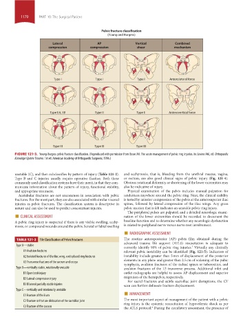
Pelvic fracture classification
(Young and Burgess)
Lateral AP Vertical Combined
compression compression shear mechanism
Type I
Type I
Type I
Type I Type I Type I Anterolateral force
Anterolateral force
Type IIA, IIB Type II Type II Anterovertical force
Type II
Type IIA, IIB
Type II
Anterovertical force
Type III Type III Type III
FIGURE 121-3. Young-Burgess pelvic fracture classification. (Reproduced with permission from Bosse MJ. The acute management of pelvic ring injuries. In: Levine AM, ed. Orthopaedic
Knowldge Update Trauma. 1st ed. American Academy of Orthopaedic Surgeons; 1996.)
unstable (C), and then subclassifies by pattern of injury (Table 121-2). and ecchymosis, that is, bleeding from the urethral meatus, vagina,
Type B and C injuries usually require operative fixation. Both these or rectum, are also good clinical signs of pelvic injury (Fig. 121-4).
commonly used classification systems have their merit, in that they com- Obvious rotational deformity or shortening of the lower extremities may
municate information about the pattern of injury, functional stability, also be indicative of injury.
and appropriate treatment. Physical examination of the pelvis includes manual palpation for
Acetabular fractures are not uncommon in association with pelvic tenderness anywhere around the pelvic ring. Next, the clinical stability
fractures. For the most part, they are also associated with similar visceral is tested by anterior compression of the pelvis at the anterosuperior iliac
injuries as pelvic fractures. The classification system is descriptive in spines, followed by lateral compression of the iliac wings. Any gross
nature and can also be used to predict concomitant injuries. pelvic motion that is felt indicates an unstable pelvic ring injury.
■ CLINICAL ASSESSMENT nation of the lower extremities should be recorded to document the
The peripheral pulses are palpated, and a detailed neurologic exami-
A pelvic ring injury is suspected if there is any visible swelling, ecchy- baseline function and to determine whether any neurologic dysfunction
mosis, or compound wounds around the pelvis. Scrotal or labial swelling is related to peripheral nerve versus nerve root involvement.
■ RADIOGRAPHIC ASSESSMENT
TABLE 121-2 Tile Classification of Pelvic Fractures The routine anteroposterior (AP) pelvis film obtained during the
advanced trauma life support (ATLS) resuscitation is adequate to
Type A—stable
correctly identify 90% of pelvic ring injuries. Virtually any clinically
7
A1 Avulsion fractures relevant pelvic instability can be identified (Fig. 121-5). Indicators of
A2 Isolated fractures of the iliac wing, undisplaced ring fractures instability include greater than 5 mm of displacement of the posterior
elements in any plane and greater than 2.5 cm of widening of the pubic
A3 Transverse fractures of the sacrum and coccyx
symphysis, avulsion fractures of the ischial spines or tuberosities, and
Type B—vertically stable, rotationally unstable avulsion fractures of the L5 transverse process. Additional inlet and
B1 Open book injury outlet radiographs are helpful to assess AP displacement and superior
migration of the hemipelvis, respectively.
B2 Lateral compression injury
For sacral fractures and subtle sacroiliac joint disruptions, the CT
B3 Bilateral partially stable injuries scan can further delineate fracture displacement.
Type C—vertically and rotationally unstable ■
C1 Fracture of the ilium MANAGEMENT
C2 Fracture or fracture dislocation of the sacroiliac joint The most important aspect of management of the patient with a pelvic
ring injury is the systemic resuscitation of hypovolemic shock as per
C3 Fracture of the sacrum
the ATLS protocol. During the circulatory assessment, the presence of
8
section10.indd 1170 1/20/2015 9:21:21 AM