
Page 1653 - Hall et al (2015) Principles of Critical Care-McGraw-Hill
P. 1653
1172 PART 10: The Surgical Patient
Provisional stabilization while the patient is being optimized in the
ICU includes external fixation for unstable injuries, as well as skeletal
traction on the lower extremity if there is associated vertical instability
of the pelvis. 19,20
Definitive stabilization for mechanically unstable pelvic ring injuries
usually requires fixation of both the posterior and anterior ring injuries,
provided there are no contraindications to surgery. The ideal time for this
is when the patient is systemically optimized and stable (Fig. 121-5B).
The use of external fixation and traction for routine injuries treatable
with open reduction and internal fixation is not advised 21,22 because this
usually compromises mobilization of the patient and increases the risk
of associated problems of prolonged bed rest, including pneumonia,
venous thromboembolism, and decubitus skin ulceration, as well as an
increased mortality rate.
■ COMPLICATIONS
Other complications may arise as a direct result of the pelvic ring injury.
Massive blood loss and hemodynamic instability as described earlier are
the most acute issues to be managed. Injuries to the gastrointestinal and
urologic systems, vaginal tears, and skin degloving can cause significant
problems if not managed properly in the acute setting.
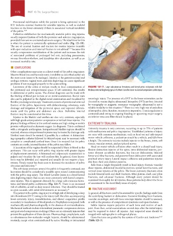
Laceration of the colon or rectum results in fecal contamination of FIGURE 121-7. Large subcutaneous hematoma and lateral pelvic ecchymosis with full-
the peritoneal and retroperitoneal space. If left untreated, this results thickness internal degloving typical of a Morel-Lavellee lesion in a lateral compression pelvic injury.
in abdominal or pelvic sepsis. The clinical diagnosis can be made with
the finding of blood per rectum or on rectal examination. Rectal tears
also can be palpated in the digital rectal examination and visualized by neurologic injury. The presence of a DVT in the lower extremities can be
flexible proctosigmoidoscopy. Treatment consists of provisional external detected by venous duplex ultrasound. Intrapelvic DVTs are best detected
fixation of the pelvis, laparotomy with defunctioning colostomy, wide by venography or magnetic resonance venography; ultrasound is not a
drainage and irrigation of the perirectal space, and repeated débride- reliable modality in this situation. There is a very high rate of embolism
25
ments until the pelvis and abdomen are clean. Definitive internal of intrapelvic clots; therefore, treatment is imperative. If anticoagulation is
fixation may be considered at this time. contraindicated owing to ongoing bleeding or upcoming major surgery,
Injuries to the bladder and urethra are also very common, especially an inferior vena cava filter should be placed.
with high-grade anteroposterior compression or vertical shear injuries. The
physical findings of blood at the urethral meatus and high-riding or mobile EXTREMITY TRAUMA
prostate with perineal ecchymosis suggest the diagnosis. This is confirmed
with a retrograde urethrogram. Intraperitoneal bladder rupture should be Extremity trauma is very common, occurring in up to 75% of patients
1
repaired, whereas extraperitoneal ruptures may be treated by drainage only. with multisystem and pelvic ring injuries. Established patterns of injury
Urethral tears should be stented, if possible, by a catheter. A defunction- are seen with common mechanisms, such as head-on and side-impact
ing suprapubic catheter followed by delayed repair may be necessary with motor vehicle collisions, a pedestrian struck by a vehicle, and falls from
complete or complicated urethral tears. Once it is ensured that the pelvic a height. The extremity injuries include injuries to the bones, joints, soft
contents are sterile, internal fixation of the pelvis may follow. tissues, vascular system, and peripheral nerves.
A laceration of the vagina should be suspected if there is blood in the Head-on motor vehicle collisions often result in a closed head injury,
perineum. This can occur with pelvic ring injuries with greater degree flexion-distraction injuries of the spine, intra-abdominal injuries, pos-
of displacement anteriorly. A bimanual and colposcopic examination to terior element acetabular fractures, hip fracture dislocations, bilateral
palpate and visualize the tear will confirm this. In general, these lacera- femur and tibia fractures, and posterior knee dislocation with associated
tions may be débrided and repaired and usually do not require a lapa- popliteal artery injury. Lateral-impact collisions and pedestrian injuries
rotomy. Timely repair has been seen to decrease the incidence of pelvic also have their own distinct patterns.
abscesses and infection. 23 Falls from a height frequently result in head injury, thoracic vascular
The soft tissue envelope around the pelvis is frequently contused; any shear injuries, abdominal visceral lacerations, spinal burst fractures, and
laceration should be considered a possible open wound communicating vertical shear injuries of the pelvis. The lower-extremity fractures often
with the pelvic ring injury. The Morel-Lavallee lesion is a closed lateral include femoral neck and shaft fractures, tibial plateau, shaft, and pilon
skin degloving injury that occurs most commonly with lateral compres- fractures, and calcaneal fractures. Although every patient should be
sion injuries. This is apparent clinically by a large subcutaneous hema- examined thoroughly for all injuries, these patterns help direct the focus
toma and lateral pelvic ecchymosis (Fig. 121-7). There is an increased of assessment to the most likely areas of injury.
risk of cellulitis, as well as deep wound infection. They should be treated ■
as open wounds, with serial débridements as necessary. 24 FRACTURE ASSESSMENT
The patient with a pelvic ring injury is at high risk of developing a deep In general, all fractures need to be assessed for specific findings aside from
vein thrombosis (DVT) owing to the injury itself, the often-accompanying the underlying fracture or dislocation. Excessive bleeding from fractures,
lower-extremity injury, immobilization, and altered coagulation profile vascular, neurologic, and soft tissue envelope injuries should be assessed,
secondary to transfusions of blood products. Prophylaxis is required, both as well as the presence of compartment syndrome and open fractures.
mechanically and pharmacologically, in the absence of contraindications. Subtle injuries require palpation of each bone and motion of each
Mechanical methods include intermittent pneumatic compression devices joint. Even then, serial examinations several days after the initial injury
and graduated compression stockings if there are no extremity injuries that may be required to detect all injuries. Any suspected areas should be
prevent the application of these devices. Pharmacologic prophylaxis, such imaged with radiographs in orthogonal planes.
as subcutaneous low-molecular-weight heparin, should be administered Open fractures are graded by the system of Gustilo and Anderson 26,27
routinely, except when contraindicated by active bleeding or intracranial (Table 121-3).
section10.indd 1172 1/20/2015 9:21:27 AM