Page 601 - Clinical Hematology_ Theory _ Procedures ( PDFDrive )
P. 601

CHAPTER 29  ■  Body Fluid Analysis                        585


                    rom the pleural, pericardial, and peritoneal cavities; synovial                                                 T ree openings in the roo  o  the  ourth ventricle, a pair

                     uid; and seminal   uid is  requently per ormed in the hema-                                               o   lateral  apertures  ( oramina  o   Luschka)  and  a  median

                   tology laboratory.                                                                                          aperture ( oramen o  Magendie), allow CSF to f ow into the

                                                                                                                               basal  cisterns  and  subarachnoid  space  o   the  spinal  cord.

                                                                                                                               From these basal cisterns, CSF migrates over the convexities
                     NOTE: This is a good time to review the de  nitions of the Key                                            toward the cerebral sinuses.

                     Terms in the Glossary and   ash cards on                                       .


                                                                                                                               Production of Cerebrospinal Fluid


                   CEREBROSPINAL FLUID                                                                                         CSF production is primarily a  unction o  the choroid plexus,


                   Anatomy and Physiology                                                                                      with a smaller proportion being derived  rom the ependy-
                                                                                                                               mal lining and perivascular spaces. T e plexus is composed

                   CSF acts as a shock absorber  or the brain and spinal cord,                                                 o  two layers: the ependyma (the lining epithelium o  the

                   circulates  nutrients,  lubricates  the  central  nervous  sys-                                             ventricle) and the pia mater. T e  olded projections o  the

                   tem (CNS), and may also contribute to the nourishment                                                       highly vascularized pia lined with epithelium are re erred

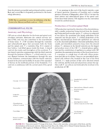
                   o  brain tissue. T e CSF circulates through the ventricles                                                  to as the choroidal epithelium. Choroidal epithelium, blood

                   and  subarachnoid  space  that  surrounds  both  the  brain                                                 vessels, and interstitial connective tissue  orm the choroid

                   and the spinal cord. T e ventricles (Fig. 29.1) consist o                                                   plexus. T e plexuses in the lateral ventricles are the largest

                    our hollow, f uid-  lled spaces inside the brain. A lateral                                                and produce most o  the CSF. T e choroid plexus epithe-

                   ventricle lies inside each hemisphere o  the cerebrum. T e                                                  lium and the endothelium o  capillaries in contact with CSF

                   two lateral ventricles communicate with the third ventricle                                                 constitute the anatomical structure o  the blood-brain barrier.

                   through the  oramen o  Monro. T e third ventricle, a nar-                                                   Te ependyma is a single layer o  cells with villous projec-

                   row  channel  between  the  hemispheres  through  the  area                                                 tions and cilia on its sur ace. Tanycytes are specialized epen-

                   o  the thalamus, communicates with the  ourth ventricle,                                                    dymal cells without cilia, located on the f oor o  the third

                   located in the pons and medulla, by means o  the aqueduct                                                   ventricle. T e main portion o  this cell is directed toward

                   o  Sylvius in the midbrain portion o  the brainstem. T is                                                   the ventricle, and the neck and tail portions contact the cap-

                   ventricle is continuous with the central canal o  the spinal                                                illary wall. T ese cells are not believed to be involved in the

                   cord.                                                                                                       production o  CSF.


                                                                                                                                                                           Longitudinal cerebral
                               Calvaria                                                                                                                                    fissures


                                                                                                                                                                           Anterior horn of lateral ventricle
                               Grey matter
                                                                                                                                                                           Head of caudate
                                                                                                                                                                           nucleus


                               Lenticular
                               nucleus


                                                                                                                                                                           Thalamus


                                                                                                                                                                           Quadrigeminal cistern

                               Choroid plexus                                                                                                                              (cistern of great cerebral vein)
                               of lateral ventricle


                               Cerebellum                                                                                                                                  Calcification in

                                                                                                                                                                           pineal body


                               Internal occipital
                               protuberance


                                     FIGURE 29.1    ransverse (axial) C   image o  the brain. Observe the ventricles, various parts o  the brain, and the
                                     choroid plexus o  the lateral ventricle. (Reprinted  rom Moore KL, Dalley AF II. Clinical Oriented Anatomy, 4th ed,

                                     Baltimore, MD: Lippincott Williams & Wilkins, 1999, with permission.)
   596   597   598   599   600   601   602   603   604   605   606