
Page 616 - Clinical Hematology_ Theory _ Procedures ( PDFDrive )
P. 616
600 PART 8 ■ Fundamentals of Hematological Analysis
serous portion o the pericardium consists o the parietal and oppose dilatation o the heart. In many circumstances, the
visceral layers. T e outer parietal layer is in contact with the restraining e ect o the pericardium is essentially ref ected
brous pericardium; the inner visceral layer, also re erred to by the mean central venous pressure. T e term cardiac
as the epicardium, is in contact with the heart and roots o tamponade is o en used to indicate a critical state o car-
the great blood vessels. T e potential space between the pari- diovascular compromise, usually with hypotension, caused
etal and visceral layers, which is lled with a small amount o by pericardial f uid under increased pressure. It is widely
f uid to reduce riction between the layers, is the pericardial accepted that any elevation o central venous pressure that is
cavity. caused by pericardial e usion constitutes cardiac tampon-
ade. T erapeutic removal o pericardial f uid, pericardiocen-
Pericardial Effusion tesis, is usually indicated i the central venous pressure rises
An abnormal accumulation o f uid in the cavity, a pericardial to approximately 10 mm Hg.
effusion, is most requently caused by damage to the lining o Pericardial e usion is usually accurately assessed by echo-
the cavity and increased capillary permeability. In addition, cardiography, but there are pit alls in the interpretation o
in acute pericarditis, inter erence with pericardial venous such studies. For example, tamponade can be produced by
and lymphatic drainage predisposes the patient to e usion localized pockets o pericardial e usion that may not be
development. evident by echocardiography, particularly i the pocket is
T e physiological unction o the normal pericardium located adjacent to the right atrium laterally. C scans and
is considered to be pericardial restraint, which tends to magnetic resonance imaging (MRI) are also accurate means
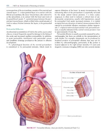
Section of heart wall
Fibrous pericardium
Parietal pericardium
Pericardial space
Epicardium
Myocardium
Endocardium
Fluid in pericardial space
FIGURE 29.7 Pericardial e usion. (Asset provided by Anatomical Chart Co.)