
Page 615 - Clinical Hematology_ Theory _ Procedures ( PDFDrive )
P. 615
CHAPTER 29 ■ Body Fluid Analysis 599
Mesothelial Cells
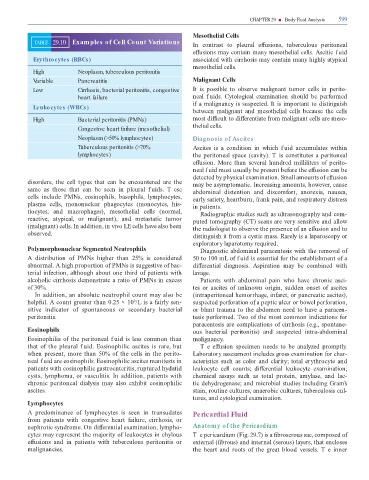
TABLE 29.10 Examples of Cell Count Variations In contrast to pleural e usions, tuberculous peritoneal
e usions may contain many mesothelial cells. Ascitic f uid
Erythrocytes (RBCs) associated with cirrhosis may contain many highly atypical
mesothelial cells.
High Neoplasm, tuberculous peritonitis
Variable Pancreatitis Malignant Cells
Low Cirrhosis, bacterial peritonitis, congestive It is possible to observe malignant tumor cells in perito-
heart failure neal f uids. Cytological examination should be per ormed
i a malignancy is suspected. It is important to distinguish
Leukocytes (WBCs)
between malignant and mesothelial cells because the cells
High Bacterial peritonitis (PMNs) most di cult to di erentiate rom malignant cells are meso-
thelial cells.
Congestive heart failure (mesothelial)
Neoplasm (>50% lymphocytes) Diagnosis of Ascites
Tuberculous peritonitis (>70% Ascites is a condition in which f uid accumulates within
lymphocytes) the peritoneal space (cavity). T is constitutes a peritoneal
e usion. More than several hundred milliliters o perito-
neal f uid must usually be present be ore the e usion can be
detected by physical examination. Small amounts o e usion
disorders, the cell types that can be encountered are the may be asymptomatic. Increasing amounts, however, cause
same as those that can be seen in pleural f uids. T ese abdominal distention and discom ort, anorexia, nausea,
cells include PMNs, eosinophils, basophils, lymphocytes, early satiety, heartburn, rank pain, and respiratory distress
plasma cells, mononuclear phagocytes (monocytes, his- in patients.
tiocytes, and macrophages), mesothelial cells (normal, Radiographic studies such as ultrasonography and com-
reactive, atypical, or malignant), and metastatic tumor puted tomography (C ) scans are very sensitive and allow
(malignant) cells. In addition, in vivo LE cells have also been the radiologist to observe the presence o an e usion and to
observed. distinguish it rom a cystic mass. Rarely is a laparoscopy or
exploratory laparotomy required.
Polymorphonuclear Segmented Neutrophils Diagnostic abdominal paracentesis with the removal o
A distribution o PMNs higher than 25% is considered 50 to 100 mL o f uid is essential or the establishment o a
abnormal. A high proportion o PMNs is suggestive o bac- di erential diagnosis. Aspiration may be combined with
terial in ection, although about one third o patients with lavage.
alcoholic cirrhosis demonstrate a ratio o PMNs in excess Patients with abdominal pain who have chronic asci-
o 30%. tes or ascites o unknown origin, sudden onset o ascites
In addition, an absolute neutrophil count may also be (intraperitoneal hemorrhage, in arct, or pancreatic ascites),
help ul. A count greater than 0.25 × 10 /L is a airly sen- suspected per oration o a peptic ulcer or bowel per oration,
9
sitive indicator o spontaneous or secondary bacterial or blunt trauma to the abdomen need to have a paracen-
peritonitis. tesis per ormed. wo o the most common indications or
paracentesis are complications o cirrhosis (e.g., spontane-
Eosinophils ous bacterial peritonitis) and suspected intra-abdominal
Eosinophilia o the peritoneal f uid is less common than malignancy.
that o the pleural f uid. Eosinophilic ascites is rare, but T e e usion specimen needs to be analyzed promptly.
when present, more than 50% o the cells in the perito- Laboratory assessment includes gross examination or char-
neal f uid are eosinophils. Eosinophilic ascites mani ests in acteristics such as color and clarity; total erythrocyte and
patients with eosinophilic gastroenteritis, ruptured hydatid leukocyte cell counts; di erential leukocyte examination;
cysts, lymphoma, or vasculitis. In addition, patients with chemical assays such as total protein, amylase, and lac-
chronic peritoneal dialysis may also exhibit eosinophilic tic dehydrogenase; and microbial studies including Gram’s
ascites. stain, routine cultures, anaerobic cultures, tuberculosis cul-
tures, and cytological examination.
Lymphocytes
A predominance o lymphocytes is seen in transudates Pericardial Fluid
rom patients with congestive heart ailure, cirrhosis, or
nephrotic syndrome. On di erential examination, lympho- Anatomy of the Pericardium
cytes may represent the majority o leukocytes in chylous T e pericardium (Fig. 29.7) is a broserous sac, composed o
e usions and in patients with tuberculous peritonitis or external ( brous) and internal (serous) layers, that encloses
malignancies. the heart and roots o the great blood vessels. T e inner