


Page 105 - Textbook of Pathology, 6th Edition
P. 105
89  CHAPTER 4


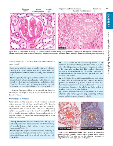
           Figure 4.12  Amyloidosis of kidney. The amyloid deposits are seen mainly in the glomerular capillary tuft. The deposits are also present in
           peritubular connective tissue producing atrophic tubules and amyloid casts in the tubular lumina, and in the arterial wall producing luminal narrowing.  Immunopathology Including Amyloidosis


           extracellular matrix and within the basement membranes of  In the glomeruli, the deposits initially appear on the
           blood vessels.                                        basement membrane of the glomerular capillaries, but
                                                                 later extend to produce luminal narrowing and distortion
            Grossly, the affected organ is usually enlarged, pale and  of the glomerular capillary tuft. This results in abnormal
            rubbery. Cut surface shows firm, waxy and translucent  increase in permeability of the glomerular capillaries to
            parenchyma which takes positive staining with the iodine  macromolecules with consequent proteinuria and
            test.
                                                                 nephrotic syndrome.
            Microscopically, the deposits of amyloid are found in the  In the tubules, the amyloid deposits likewise begin close
            extracellular locations, initially in the walls of small blood  to the tubular epithelial basement membrane. Subse-
            vessels producing microscopic changes and effects, while  quently, the deposits may extend further outwards into
            later the deposits are in large amounts causing      the intertubular connective tissue, and inwards to produce
            macroscopic changes and effects of pressure atrophy.  degenerative changes in the tubular epithelial cells and
                                                                 amyloid casts in the tubular lumina.
              Based on these general features of amyloidosis, the salient
           pathologic findings of major organ involvements are      Vascular involvement affects chiefly the walls of small
           described below.                                      arterioles and venules, producing narrowing of their
                                                                 lumina and consequent ischaemic effects.
           Amyloidosis of Kidneys
           Amyloidosis of the kidneys is most common and most
           serious because of ill-effects on renal function. The deposits
           in the kidneys are found in most cases of secondary
           amyloidosis and in about one-third cases of primary
           amyloidosis. Amyloidosis of the kidney accounts for about
           20% of deaths from amyloidosis. Even small quantities of
           amyloid deposits in the glomeruli can cause proteinuria and
           nephrotic syndrome.

            Grossly, the kidneys may be normal-sized, enlarged or
            terminally contracted due to ischaemic effect of narrowing
            of vascular lumina. Cut surface is pale, waxy and
            translucent (Fig. 4.11).
            Microscopically, amyloid deposition occurs primarily in
            the glomeruli, though it may involve peritubular   Figure 4.13  Amyloidosis kidney, Congo red stain. A, The amyloid
            interstitial tissue and the walls of arterioles as well  deposits are seen mainly in the glomerular capillary tuft stained red-pink
            (Fig. 4.12):                                       (Congophilia). B, Viewing the same under polarising microscopy, the
                                                               congophilic areas show apple-green birefringence.

