


Page 104 - Textbook of Pathology, 6th Edition
P. 104
88 Metachromatic stains employed are rosaniline dyes such as  produce symptoms and may even cause death. The diagnosis
           methyl violet and crystal violet which impart  rose-pink  of amyloid disease can be made from the following
           colouration to amyloid deposits. However, small amounts  investigations:
           of amyloid are missed, mucins also have metachromasia and  1. BIOPSY EXAMINATION. Histologic examination of
           that aqueous mountants are required for seeing the  biopsy material is the commonest and confirmatory method
           preparation.  Therefore, this method has low sensitivity and  for diagnosis in a suspected case of amyloidosis. Biopsy of
           lacks specificity.                                  an obviously affected organ is likely to offer the best results
           4. CONGO RED AND POLARISED LIGHT. All types of      e.g. kidney biopsy in a case of dialysis, sural nerve biopsy in
           amyloid have affinity for Congo red stain; therefore this  familial polyneuropathy. In systemic amyloidosis, renal
     SECTION I
           method is used for confirmation of amyloid of all types. The  biopsy provides the best detection rate, but rectal biopsy also
           stain may be used on both gross specimens and microscopic  has a good pick up rate. However, gingiva and skin biopsy
           sections; amyloid of all types stains  pink  red colour. If the  have poor result. Currently, fine needle aspiration of
           stained section is viewed in polarised light, the amyloid  abdominal subcutaneous fat followed by Congo red staining
           characteristically shows apple-green birefringence due to cross-  and polarising microscopic examination for confirmation has
           β-pleated sheet configuration of amyloid fibrils. The stain  become an acceptable simple and useful technique with
           can also be used to distinguish between AL and AA amyloid  excellent result.
           (primary and secondary amyloid respectively). After prior  2. IN VIVO CONGO RED TEST. A known quantity of
           treatment with permanganate or trypsin on the section,  Congo red dye may be injected intravenously in living
           Congo red stain is repeated—in the case of primary amyloid  patient. If amyloidosis is present, the dye gets bound to
           (AL amyloid), the Congo red positivity (congophilia)  amyloid deposits and its levels in blood rapidly decline. The
           persists,* while it turns negative for Congo red in secondary  test is, however, not popular due to the risk of anaphylaxis
           amyloid (AA amyloid). Congo red dye can also be used as  to the injected dye.
           an in vivo test (described below).
                                                               3. OTHER TESTS.  A few other tests which are not
           5. FLUORESCENT STAINS. Fluorescent stain thioflavin-  diagnostic but are supportive of amyloid disease are protein
           T binds to amyloid and fluoresce yellow under ultraviolet  electrophoresis, immunoelectrophoresis of urine and serum,
           light i.e. amyloid emits secondary fluorescence. Thioflavin-  and bone marrow aspiration.
           S is less specific.
           6. IMMUNOHISTOCHEMISTRY. More recently, type of     MORPHOLOGIC FEATURES OF
           amyloid can be classified by immunohistochemical stains.  AMYLOIDOSIS OF ORGANS
     General Pathology and Basic Techniques
           Various antibody stains against the specific antigenic  Although amyloidosis of different organs shows variation
           protein types of amyloid are commercially available.  in morphologic pattern, some features are applicable in
           However, most useful in confirmation for presence of  general to most of the involved organs.
           amyloid of any type is anti-AP stain; others for determining
           the biochemical type of amyloid include anti-AA, anti-lambda  Sites of Amyloid Deposits. In general, amyloid proteins get
           (λ), anti- kappa (κ) antibody stains etc.           filtered from blood across the basement membrane of
                                                               vascular capillaries into extravascular spaces.  Thus, most
           7. NON-SPECIFIC STAINS. A few other stains have been  commonly amyloid deposits appear at the contacts between
           described for amyloid at different times but they lack  the vascular spaces and parenchymal cells, in the
           specificity. These are as under:
           i) Standard toluidine blue: This method gives orthochromatic
           blue colour to amyloid which under polarising microscopy
           produces dark red birefringence. However, there are false
           positive as well as false negative results; hence not
           recommended.
           ii) Alcian blue:  It imparts  blue-green colour to amyloid
           positive areas and is used for mucopolysaccharide content
           in amyloid but uptake of dye is poor and variable.
           iii) Periodic acid Schiff (PAS): It is used for demonstration
           of  carbohydrate content of amyloid but shows variable
           positivity and is not specific.

           DIAGNOSIS OF AMYLOIDOSIS
           Amyloidosis may be detected as an unsuspected morpho-
           logic finding in a case, or the changes may be severe so as to

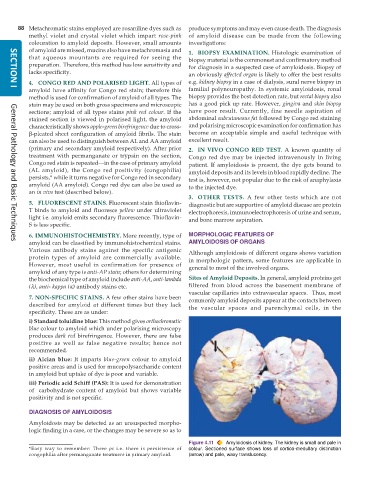
                                                               Figure 4.11  Amyloidosis of kidney. The kidney is small and pale in
           *Easy way to remember: Three  ps  i.e. there is  persistence of  colour. Sectioned surface shows loss of cortico-medullary distinction
           congophilia after permanganate treatment in primary amyloid.  (arrow) and pale, waxy translucency.

