


Page 106 - Textbook of Pathology, 6th Edition
P. 106
90


     SECTION I


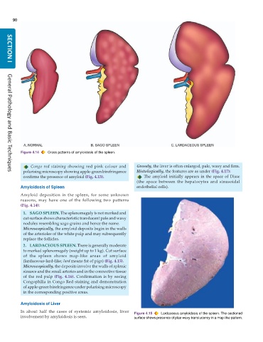
           Figure 4.14  Gross patterns of amyloidosis of the spleen.

               Congo red staining  showing red pink colour and   Grossly, the liver is often enlarged, pale, waxy and firm.
            polarising microscopy showing apple-green birefringence  Histologically, the features are as under (Fig. 4.17):
     General Pathology and Basic Techniques
            confirms the presence of amyloid (Fig. 4.13).           The amyloid initially appears in the space of Disse
                                                                 (the space between the hepatocytes and sinusoidal
           Amyloidosis of Spleen                                 endothelial cells).
           Amyloid deposition in the spleen, for some unknown
           reasons, may have one of the following two patterns
           (Fig. 4.14):
            1. SAGO SPLEEN. The splenomegaly is not marked and
            cut surface shows characteristic translucent pale and waxy
            nodules resembling sago grains and hence the name.
            Microscopically, the amyloid deposits begin in the walls
            of the arterioles of the white pulp and may subsequently
            replace the follicles.
            2. LARDACEOUS SPLEEN. There is generally moderate
            to marked splenomegaly (weight up to 1 kg). Cut surface
            of the spleen shows map-like areas of amyloid
            (lardaceous-lard-like; lard means fat of pigs) (Fig. 4.15).
            Microscopically, the deposits involve the walls of splenic
            sinuses and the small arteries and in the connective tissue
            of the red pulp (Fig. 4.16). Confirmation is by seeing
            Congophilia in Congo Red staining and demonstration
            of apple-green birefringence under polarising microscopy
            in the corresponding positive areas.

           Amyloidosis of Liver
           In about half the cases of systemic amyloidosis, liver  Figure 4.15  Lardaceous amyloidosis of the spleen. The sectioned
           involvement by amyloidosis is seen.                 surface shows presence of plae waxy translucency in a map-like pattern.

