


Page 136 - Textbook of Pathology, 6th Edition
P. 136
120 pulmonary veins and paradoxical arterial emboli from the
TABLE 5.7: Important Types of Embolism.
systemic venous circulation.
Type Common Origin The effects of arterial emboli depend upon their size, site
1. Pulmonary embolism Veins of lower legs of lodgement, and adequacy of collateral circulation. If the
2. Systemic embolism Left ventricle (arterial) vascular occlusion occurs, the following ill-effects may result:
3. Fat embolism Trauma to bones/soft tissues i) Infarction of the organ or its affected part e.g. ischaemic
4. Air embolism Venous: head and neck necrosis in the lower limbs (70-75%), spleen, kidneys, brain,
operations, obstetrical trauma intestine.
Arterial: cardiothoracic ii) Gangrene following infarction in the lower limbs if the
SECTION I
surgery, angiography collateral circulation is inadequate.
5. Decompression Descent: divers
sickness Ascent: unpressurised flight iii) Arteritis and mycotic aneurysm formation from bacterial
6. Amniotic fluid embolism Components of amniotic fluid endocarditis.
7. Atheroembolism Atheromatous plaques iv) Myocardial infarction may occur following coronary
8. Tumour embolism Tumour fragments embolism.
v) Sudden death may result from coronary embolism or
embolism in the middle cerebral artery.
ii) Retrograde embolus. An embolus which travels against the
flow of blood is called retrograde embolus e.g. metastatic Venous thromboembolism. Venous emboli may arise from
deposits in the spine from carcinoma prostate. The spread the following sources:
occurs by retrograde embolism through intraspinal veins i) Thrombi in the veins of the lower legs are the most
which carry tumour emboli from large thoracic and common cause of venous emboli.
abdominal veins due to increased pressure in body cavities ii) Thrombi in the pelvic veins.
e.g. during coughing or straining. iii) Thrombi in the veins of the upper limbs.
Some of the important types of embolism are tabulated iv) Thrombosis in cavernous sinus of the brain.
in Table 5.7 and described below: v) Thrombi in the right side of heart.
The most significant effect of venous embolism is
Thromboembolism
obstruction of pulmonary arterial circulation leading to
A detached thrombus or part of thrombus constitutes the pulmonary embolism described below.
most common type of embolism. These may arise in the
General Pathology and Basic Techniques
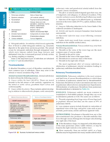
arterial or venous circulation (Fig. 5.24): Pulmonary Thromboembolism
Arterial (systemic) thromboembolism. Arterial emboli may DEFINITION. Pulmonary embolism is the most common
be derived from the following sources: and fatal form of venous thromboembolism in which there
A. Causes within the heart (80-85%): These are mural is occlusion of pulmonary arterial tree by thromboemboli.
thrombi in the left atrium or left ventricle, vegetations on Pulmonary thrombosis as such is uncommon and may occur
the mitral or aortic valves, prosthetic heart valves and in pulmonary atherosclerosis and pulmonary hypertension.
cardiomyopathy. Differentiation of pulmonary thrombosis from pulmonary
B. Causes within the arteries: These include emboli develop- thromboembolism is tabulated in Table 5.8.
ing in relation to atherosclerotic plaques, aortic aneurysms,
ETIOLOGY. Pulmonary emboli are more common in
hospitalised or bed-ridden patients, though they can occur
in ambulatory patients as well. The causes are as follows:
i) Thrombi originating from large veins of lower legs (such
as popliteal, femoral and iliac) are the cause in 95% of
pulmonary emboli.
ii) Less common sources include thrombi in varicosities of
superficial veins of the legs, and pelvic veins such as peri-
prostatic, periovarian, uterine and broad ligament veins.
PATHOGENESIS. Detachment of thrombi from any of the
above-mentioned sites produces a thrombo-embolus that
flows through venous drainage into the larger veins draining
into right side of the heart.
If the thrombus is large, it is impacted at the bifurcation
of the main pulmonary artery (saddle embolus), or may be
found in the right ventricle or its outflow tract.
More commonly, there are multiple emboli, or a large
Figure 5.24 Sources of arterial and venous emboli. embolus may be fragmented into many smaller emboli which

