


Page 134 - Textbook of Pathology, 6th Edition
P. 134
118
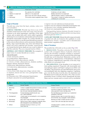
TABLE 5.5: Distinguishing Features of Antemortem Thrombi and Postmortem Clots.
Feature Antemortem Thrombi Postmortem Clots
1. Gross Dry, granular, firm and friable Gelatinous, soft and rubbery
2. Relation to vessel wall Adherent to the vessel wall Weakly attached to the vessel wall
3. Shape May or may not fit their vascular contours Take the shape of vessel or its bifurcation
4. Microscopy The surface contains apparent lines of Zahn The surface is ‘chicken fat’ yellow covering the
underlying red ‘currant jelly’
SECTION I
Origin of Thrombi v) Superior vena cava: infections in head and neck.
Thrombi may arise from the heart, arteries, veins or in vi) Inferior vena cava: extension of thrombus from hepatic vein.
microcirculation. vii) Mesenteric veins: volvulus, intestinal obstruction.
viii) Renal vein: renal amyloidosis.
CARDIAC THROMBI. Thrombi may form in any of the
chambers of the heart and on the valve cusps. They are more Distinguishing features between thrombi formed in
common in the atrial appendages, especially of the right rapidly-flowing arterial circulation and slow-moving venous
atrium, and on mitral and aortic valves called vegetations blood are given in Table 5.6.
which may be seen in infective endocarditis and non-bacterial CAPILLARY THROMBI. Minute thrombi composed mainly
thrombotic endocarditis (Chapter 16). Cardiac thrombi are of packed red cells are formed in the capillaries in acute
mural (non-occlusive) as are the mural thrombi encountered inflammatory lesions, vasculitis and in disseminated
in the aorta in atherosclerosis and in aneurysmal dilatations. intravascular coagulation (DIC).
Rarely, large round thrombus may form and obstruct the
mitral valve and is called ball-valve thrombus. Agonal thrombi Fate of Thrombus
are formed shortly before death and may occur in either or The possible fate of thrombi can be as under (Fig. 5.23):
both the ventricles. They are composed mainly of fibrin.
1. RESOLUTION. Thrombus activates the fibrinolytic
ARTERIAL AND VENOUS THROMBI. The examples of system with consequent release of plasmin which may
major forms of vascular thrombi are as under:
dissolve the thrombus completely resulting in resolution.
Arterial thrombi: Usually, lysis is complete in small venous thrombi while large
i) Aorta: aneurysms, arteritis. thrombi may not be dissolved. Fibrinolytic activity can be
General Pathology and Basic Techniques
ii) Coronary arteries: atherosclerosis. accentuated by administration of thrombolytic substances
iii) Mesenteric artery: atherosclerosis, arteritis. (e.g. urokinase, streptokinase), especially in the early stage
iv) Arteries of limbs: atherosclerosis, diabetes mellitus, when fibrin is in monomeric form.
Buerger’s disease, Raynaud’s disease. 2. ORGANISATION. If the thrombus is not removed, it
v) Renal artery: atherosclerosis, arteritis. starts getting organised. Phagocytic cells (neutrophils and
vi) Cerebral artery: atherosclerosis, vasculitis.
macrophages) appear and begin to phagocytose fibrin and
Venous thrombi: cell debris. The proteolytic enzymes liberated by leucocytes
i) Veins of lower limbs: deep veins of legs, varicose veins. and endothelial cells start digesting coagulum. Capillaries
ii) Popliteal, femoral and iliac veins: postoperative stage, grow into the thrombus from the site of its attachment and
postpartum. fibroblasts start invading the thrombus. Thus, fibrovascular
iii) Pulmonary veins: CHF, pulmonary hypertension. granulation tissue is formed which subsequently becomes
iv) Hepatic and portal vein: portal hypertension. dense and less vascular and is covered over by endothelial
TABLE 5.6: Distinguishing Features of Arterial and Venous Thrombi.
Feature Arterial Thrombi Venous Thrombi
1. Blood flow Formed in rapidly-flowing blood of arteries and heart Formed in slow-moving blood in veins
2. Sites Common in aorta, coronary, cerebral, Common in superficial varicose veins, deep leg
iliac, femoral, renal and mesenteric arteries veins, popliteal, femoral and iliac veins
3. Thrombogenesis Formed following endothelial cell injury Formed following venous stasis e.g. in abdominal
e.g. in atherosclerosis operations, child-birth
4. Development Usually mural, not occluding the lumen completely, Usually occlusive, take the cast of the vessel in
may propagate which formed, may propagate in both directions
5. Macroscopy Grey-white, friable with lines of Zahn on surface Red-blue with fibrin strands and lines of Zahn
6. Microscopy Distinct lines of Zahn composed of platelets, fibrin Lines of Zahn with more abundant red cells
with entangled red and white blood cells
7. Effects Ischaemia leading to infarcts e.g. in the Thromboembolism, oedema, skin ulcers, poor
heart, brain etc wound healing

