


Page 188 - Textbook of Pathology, 6th Edition
P. 188
172                                                      external callus is cleared away, compact bone (cortex) is
                                                               formed in place of intermediate callus and the bone marrow
                                                               cavity develops in internal callus.
                                                               COMPLICATIONS OF FRACTURE HEALING. These are
                                                               as under:
                                                               1. Fibrous union may result instead of osseous union if the
                                                               immobilisation of fractured bone is not done. Occasionally,
                                                               a false joint may develop at the fracture site (pseudo-
     SECTION I
                                                               arthrosis).
                                                               2. Non-union may result if some soft tissue is interposed
                                                               between the fractured ends.
                                                               3. Delayed union may occur from causes of delayed wound
                                                               healing in general such as infection, inadequate blood supply,
                                                               poor nutrition, movement and old age.

                                                               Healing of Nervous Tissue
                                                               CENTRAL NERVOUS SYSTEM.  The nerve cells of the
                                                               brain, spinal cord and ganglia once destroyed are not
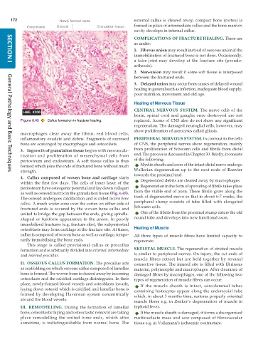
           Figure 6.45  Callus formation in fracture healing.  replaced. Axons of CNS also do not show any significant
                                                               regeneration. The damaged neuroglial cells, however, may
                                                               show proliferation of astrocytes called gliosis.
           macrophages clear away the fibrin, red blood cells,
           inflammatory exudate and debris. Fragments of necrosed  PERIPHERAL NERVOUS SYSTEM. In contrast to the cells
           bone are scavenged by macrophages and osteoclasts.  of CNS, the peripheral nerves show regeneration, mainly
           3. Ingrowth of granulation tissue begins with neovascula-  from proliferation of Schwann cells and fibrils from distal
           risation and proliferation of mesenchymal cells from  end. The process is discussed in Chapter 30. Briefly, it consists
           periosteum and endosteum. A soft tissue callus is thus  of the following:
           formed which joins the ends of fractured bone without much  Myelin sheath and axon of the intact distal nerve undergo
           strength.                                           Wallerian degeneration up to the next node of Ranvier
     General Pathology and Basic Techniques
           4. Callus composed of woven bone and cartilage starts  towards the proximal end.
           within the first few days. The cells of inner layer of the  Degenerated debris are cleared away by macrophages.
           periosteum have osteogenic potential and lay down collagen  Regeneration in the form of sprouting of fibrils takes place
           as well as osteoid matrix in the granulation tissue (Fig. 6.45).  from the viable end of axon. These fibrils grow along the
                                                               track of degenerated nerve so that in about 6-7 weeks, the
           The osteoid undergoes calcification and is called woven bone
           callus. A much wider zone over the cortex on either side of  peripheral stump consists of tube filled with elongated
           fractured ends is covered by the woven bone callus and  Schwann cells.
           united to bridge the gap between the ends, giving spindle-  One of the fibrils from the proximal stump enters the old
           shaped or fusiform appearance to the union. In poorly  neural tube and develops into new functional axon.
           immobilised fractures (e.g. fracture ribs), the subperiosteal
           osteoblasts may form cartilage at the fracture site. At times,  Healing of Muscle
           callus is composed of woven bone as well as cartilage, tempo-  All three types of muscle fibres have limited capacity to
           rarily immobilising the bone ends.                  regenerate.
              This stage is called provisional callus or procallus
           formation and is arbitrarily divided into external, intermediate  SKELETAL MUSCLE. The regeneration of striated muscle
           and internal procallus.                             is similar to peripheral nerves. On injury, the cut ends of
                                                               muscle fibres retract but are held together by stromal
           II. OSSEOUS CALLUS FORMATION. The procallus acts    connective tissue. The injured site is filled with fibrinous
           as scaffolding on which osseous callus composed of lamellar  material, polymorphs and macrophages. After clearance of
           bone is formed. The woven bone is cleared away by incoming  damaged fibres by macrophages, one of the following two
           osteoclasts and the calcified cartilage disintegrates. In their  types of regeneration of muscle fibres can occur:
           place, newly-formed blood vessels and osteoblasts invade,  If the muscle sheath is intact, sarcolemmal tubes
           laying down osteoid which is calcified and lamellar bone is  containing histiocytes appear along the endomysial tube
           formed by developing Haversian system concentrically  which, in about 3 months time, restores properly oriented
           around the blood vessels.
                                                               muscle fibres e.g. in Zenker’s degeneration of muscle in
           III.   REMODELLING.  During the formation of lamellar  typhoid fever.
           bone, osteoblastic laying and osteoclastic removal are taking  If the muscle sheath is damaged, it forms a disorganised
           place remodelling the united bone ends, which after  multinucleate mass and scar composed of fibrovascular
           sometime, is indistinguishable from normal bone. The  tissue e.g. in Volkmann’s ischaemic contracture.

