


Page 183 - Textbook of Pathology, 6th Edition
P. 183
167


                                                                                                                      CHAPTER 6


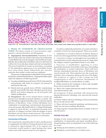
           Figure 6.41  Active granulation tissue has inflammatory cell infiltrate, newly formed blood vessels and young fibrous tissue in loose matrix.  Inflammation and Healing

           3. PHASE OF INGROWTH OF GRANULATION                    In order to explain the mechanism of wound contraction,
           TISSUE. This phase consists of 2 main processes: angio-  a number of factors have been proposed. These are as under:
           genesis or neovascularisation, and fibrogenesis.    1. Dehydration as a result of removal of fluid by drying of
           i) Angiogenesis (neovascularisation). Formation of new  wound was first suggested but without being substantiated.
           blood vessels at the site of injury takes place by proliferation  2. Contraction of collagen was thought to be responsible for
           of endothelial cells from the margins of severed blood vessels.  contraction but wound contraction proceeds at a stage when
           Initially, the proliferated endothelial cells are solid buds but  the collagen content of granulation tissue is very small.
           within a few hours develop a lumen and start carrying blood.  3. Discovery of  myofibroblasts appearing in active
           The newly formed blood vessels are more leaky, accounting  granulation tissue has resolved the controversy surrounding
           for the oedematous appearance of new granulation tissue.  the mechanism of wound contraction. These cells have
           Soon, these blood vessels differentiate into muscular  features intermediate between those of fibroblasts and
           arterioles, thin-walled venules and true capillaries.  smooth muscle cells. Their migration into the wound area
              The process of angiogenesis is stimulated with proteolytic  and their active contraction decreases the size of the defect.
           destruction of basement membrane. Angiogenesis takes place  The evidences in support of this concept are both
           under the influence of following factors:
                                                               morphological as well as functional characteristics of
           a) Vascular endothelial growth factor (VEGF) elaborated by  modified fibroblasts or myofibroblasts as under:
              mesenchymal cells while its receptors are present in  i) Fibrils present in the cytoplasm of these cells resemble
              endothelial cells only.                          those seen in smooth muscle cells.
           b) Platelet-derived growth factor (PDGF), transforming  ii) These cells contain actin-myosin similar to that found in
              growth factor-β (TGF-β), basic fibroblast growth factor  non-striated muscle cells.
              (bFGF) and surface integrins are all associated with  iii) Cytoplasm of these modified cells demonstrates
              cellular proliferation.                          immunofluorescent labelling with anti-smooth muscle
           ii) Fibrogenesis. The newly formed blood vessels are  antibodies.
           present in an amorphous ground substance or matrix. The  iv) Nuclei of these cells have infoldings of nuclear membrane
           new fibroblasts originate from fibrocytes as well as by mitotic  like in smooth muscle cells.
           division of fibroblasts. Some of these fibroblasts have
           combination of morphologic and functional characteristics  v) These cells have basement membrane and desmosomes
           of smooth muscle cells (myofibroblasts). Collagen fibrils begin  which are not seen in ordinary fibroblasts.
           to appear by about 6th day. As maturation proceeds, more  vi) Drug response of granulation tissue is similar to that of
           and more of collagen is formed while the number of active  smooth muscle.
           fibroblasts and new blood vessels decreases. This results in
           formation of inactive looking scar known as cicatrisation.  WOUND HEALING

           Contraction of Wounds                               Healing of skin wounds provides a classical example of
                                                               combination of regeneration and repair described above.
           The wound starts contracting after 2-3 days and the process
           is completed by the 14th day. During this period, the wound  Wound healing can be accomplished in one of the following
           is reduced by approximately 80% of its original size.  two ways:
           Contracted wound results in rapid healing since lesser  Healing by first intention (primary union)
           surface area of the injured tissue has to be replaced.  Healing by second intention (secondary union).

