


Page 184 - Textbook of Pathology, 6th Edition
P. 184
168


     SECTION I


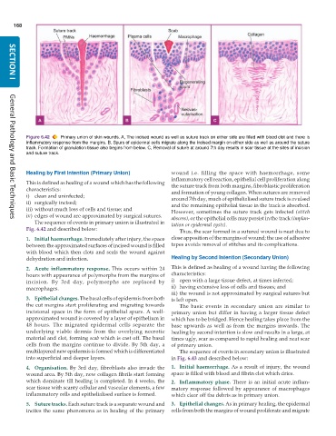
           Figure 6.42  Primary union of skin wounds. A, The incised wound as well as suture track on either side are filled with blood clot and there is
           inflammatory response from the margins. B, Spurs of epidermal cells migrate along the incised margin on either side as well as around the suture
           track. Formation of granulation tissue also begins from below. C, Removal of suture at around 7th day results in scar tissue at the sites of incision
           and suture track.


           Healing by First Intention (Primary Union)          wound i.e. filling the space with haemorrhage, some
                                                               inflammatory cell reaction, epithelial cell proliferation along
           This is defined as healing of a wound which has the following  the suture track from both margins, fibroblastic proliferation
           characteristics:                                    and formation of young collagen. When sutures are removed
           i) clean and uninfected;                            around 7th day, much of epithelialised suture track is avulsed
           ii) surgically incised;                             and the remaining epithelial tissue in the track is absorbed.
           iii) without much loss of cells and tissue; and
           iv) edges of wound are approximated by surgical sutures.  However, sometimes the suture track gets infected (stitch
     General Pathology and Basic Techniques
              The sequence of events in primary union is illustrated in  abscess), or the epithelial cells may persist in the track (implan-
                                                               tation or epidermal cysts).
           Fig. 6.42 and described below:                         Thus, the scar formed in a sutured wound is neat due to
           1. Initial haemorrhage. Immediately after injury, the space  close apposition of the margins of wound; the use of adhesive
           between the approximated surfaces of incised wound is filled  tapes avoids removal of stitches and its complications.
           with blood which then clots and seals the wound against
           dehydration and infection.                          Healing by Second Intention (Secondary Union)
           2. Acute inflammatory response. This occurs within 24  This is defined as healing of a wound having the following
           hours with appearance of polymorphs from the margins of  characteristics:
           incision. By 3rd day, polymorphs are replaced by    i) open with a large tissue defect, at times infected;
           macrophages.                                        ii) having extensive loss of cells and tissues; and
                                                               iii) the wound is not approximated by surgical sutures but
           3. Epithelial changes. The basal cells of epidermis from both  is left open.
           the cut margins start proliferating and migrating towards  The basic events in secondary union are similar to
           incisional space in the form of epithelial spurs. A well-  primary union but differ in having a larger tissue defect
           approximated wound is covered by a layer of epithelium in  which has to be bridged. Hence healing takes place from the
           48 hours. The migrated epidermal cells separate the  base upwards as well as from the margins inwards. The
           underlying viable dermis from the overlying necrotic  healing by second intention is slow and results in a large, at
           material and clot, forming scab which is cast off. The basal  times ugly, scar as compared to rapid healing and neat scar
           cells from the margins continue to divide. By 5th day, a  of primary union.
           multilayered new epidermis is formed which is differentiated  The sequence of events in secondary union is illustrated
           into superficial and deeper layers.                 in Fig. 6.43 and described below:

           4. Organisation. By 3rd day, fibroblasts also invade the  1. Initial haemorrhage. As a result of injury, the wound
           wound area. By 5th day, new collagen fibrils start forming  space is filled with blood and fibrin clot which dries.
           which dominate till healing is completed. In 4 weeks, the  2. Inflammatory phase. There is an initial acute inflam-
           scar tissue with scanty cellular and vascular elements, a few  matory response followed by appearance of macrophages
           inflammatory cells and epithelialised surface is formed.  which clear off the debris as in primary union.
           5. Suture tracks. Each suture track is a separate wound and  3. Epithelial changes. As in primary healing, the epidermal
           incites the same phenomena as in healing of the primary  cells from both the margins of wound proliferate and migrate

