


Page 185 - Textbook of Pathology, 6th Edition
P. 185
169
CHAPTER 6
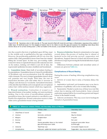
Figure 6.43 Secondary union of skin wounds. A, The open wound is filled with blood clot and there is inflammatory response at the junction of Inflammation and Healing
viable tissue. B, Epithelial spurs from the margins of wound meet in the middle to cover the gap and separate the underlying viable tissue from
necrotic tissue at the surface forming scab. C, After contraction of the wound, a scar smaller than the original wound is left.
into the wound in the form of epithelial spurs till they meet 6. Presence of infection. Bacterial contamination of an open
in the middle and re-epithelialise the gap completely. wound delays the process of healing due to release of
However, the proliferating epithelial cells do not cover the bacterial toxins that provoke necrosis, suppuration and
surface fully until granulation tissue from base has started thrombosis. Surgical removal of dead and necrosed tissue,
filling the wound space. In this way, pre-existing viable debridement, helps in preventing the bacterial infection of open
connective tissue is separated from necrotic material and clot wounds.
on the surface, forming scab which is cast off. In time, the Differences between primary and secondary union of
regenerated epidermis becomes stratified and keratinised. wounds are given in Table 6.6.
4. Granulation tissue. Main bulk of secondary healing is
by granulations. Granulation tissue is formed by proliferation Complications of Wound Healing
of fibroblasts and neovascularisation from the adjoining During the course of healing, following complications may
viable elements. The newly-formed granulation tissue is deep occur:
red, granular and very fragile. With time, the scar on
maturation becomes pale and white due to increase in 1. Infection of wound due to entry of bacteria delays the
collagen and decrease in vascularity. Specialised structures healing.
of the skin like hair follicles and sweat glands are not replaced 2. Implantation (epidermal) cyst formation may occur due to
unless their viable residues remain which may regenerate. persistence of epithelial cells in the wound after healing.
3. Pigmentation. Healed wounds may at times have rust-like
5. Wound contraction. Contraction of wound is an colour due to staining with haemosiderin. Some coloured
important feature of secondary healing, not seen in primary particulate material left in the wound may persist and impart
healing. Due to the action of myofibroblasts present in colour to the healed wound.
granulation tissue, the wound contracts to one-third to one-
fourth of its original size. Wound contraction occurs at a time 4. Deficient scar formation. This may occur due to inadequate
when active granulation tissue is being formed. formation of granulation tissue.
TABLE 6.6: Differences between Primary and Secondary Union of Wounds.
Feature Primary Union Secondary Union
1. Cleanliness of wound Clean Unclean
2. Infection Generally uninfected May be infected
3. Margins Surgical clean Irregular
4. Sutures Used Not used
5. Healing Scanty granulation tissue at the incised Exuberant granulation tissue
gap and along suture tracks to fill the gap
6. Outcome Neat linear scar Contracted irregular wound
7. Complications Infrequent, epidermal inclusion cyst formation Suppuration, may require debridement

