


Page 198 - Textbook of Pathology, 6th Edition
P. 198
182 MYCETOMA
           Mycetoma is a chronic suppurative infection involving a
           limb, shoulder or other tissues and is characterised by
           draining sinuses. The material discharged from the sinuses
           is in the form of grains consisting of colonies of fungi or
           bacteria. Mycetomas are of 2 main types:
              Mycetomas caused by actinomyces (higher bacteria)
           comprising about 60% of cases (page 163).
     SECTION I
              Eumycetomas caused by true fungi comprising the
           remaining 40% of the cases.
              Most common fungi causative for eumycetoma are
           Madurella mycetomatis or Madurella grisea, both causing black
           granules from discharging sinuses. Eumycetomas are
           particularly common in Northern and tropical Africa,
           Southern Asia and tropical America. The organisms are
           inoculated directly from soil into barefeet, from carrying of
           contaminated sacks on the shoulders, and into the hands from
           infected vegetation.
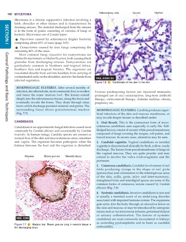
                                                               Figure 7.8  Candidiasis of the ulcer in the skin.
            MORPHOLOGIC FEATURES. After several months of
            infection, the affected site, most commonly foot, is swollen  Various predisposing factors are: impaired immunity,
            and hence the name ‘madura foot’. The lesions extend  prolonged use of oral contraceptives, long-term antibiotic
            deeply into the subcutaneous tissues, along the fascia and  therapy, corticosteroid therapy, diabetes mellitus, obesity,
            eventually invade the bones. They drain through sinus  pregnancy etc.
            tracts which discharge purulent material and grains. The
            surrounding tissue shows granulomatous reaction      MORPHOLOGIC FEATURES. Candida produces super-
            (Fig. 7.7).                                          ficial infections of the skin and mucous membranes, or
                                                                 may invade deeper tissues as described under:
           CANDIDIASIS                                           1. Oral thrush. This is the commonest form of muco-
     General Pathology and Basic Techniques
           Candidiasis is an opportunistic fungal infection caused most  cutaneous candidiasis seen especially in early life. Full-
           commonly by Candida albicans and occasionally by Candida  fledged lesions consist of creamy white pseudomembrane
           tropicalis. In human beings, Candida species are present as  composed of fungi covering the tongue, soft palate, and
           normal flora of the skin and mucocutaneous areas, intestines  buccal mucosa. In severe cases, ulceration may be seen.
           and vagina. The organism becomes pathogenic when the  2. Candidal vaginitis. Vaginal candidiasis or monilial
           balance between the host and the organism is disturbed.  vaginitis is characterised clinically by thick, yellow, curdy
                                                                 discharge. The lesions form pseudomembrane of fungi on
                                                                 the vaginal mucosa. They are quite pruritic and may
                                                                 extend to involve the vulva (vulvovaginitis) and the
                                                                 perineum.
                                                                 3. Cutaneous candidiasis. Candidal involvement of nail
                                                                 folds producing change in the shape of nail plate
                                                                 (paronychia) and colonisation in the intertriginous areas
                                                                 of the skin, axilla, groin, infra- and inter-mammary,
                                                                 intergluteal folds and interdigital spaces are some of the
                                                                 common forms of cutaneous lesions caused by Candida
                                                                 albicans (Fig. 7.8).
                                                                 4. Systemic candidiasis. Invasive candidiasis is rare and
                                                                 is usually a terminal event of an underlying disorder
                                                                 associated with impaired immune system. The organisms
                                                                 gain entry into the body through an ulcerative lesion on
                                                                 the skin and mucosa or may be introduced by iatrogenic
                                                                 means such as via intravenous infusion, peritoneal dialysis
                                                                 or urinary catheterisation. The lesions of systemic
                                                                 candidiasis are most commonly encountered in kidneys
                                                                 as ascending pyelonephritis and in heart as candidal
           Figure 7.7  Madura foot. Brown granule lying in necrotic tissue in  endocarditis.
           the discharging sinus.

