


Page 216 - Textbook of Pathology, 6th Edition
P. 216
200                                                      discontinuous secondary tumour mass/masses are formed
                                                               at the site of lodgement. Metastasis and invasiveness are the
                                                               two most important features to distinguish malignant from benign
                                                               tumours:  benign tumours do not metastasise while all the
                                                               malignant tumours with a few exceptions like gliomas of the
                                                               central nervous system and basal cell carcinoma of the skin,
                                                               can metastasise. Generally, larger, more aggressive and
                                                               rapidly-growing tumours are more likely to metastasise but
                                                               there are numerous exceptions. About one-third of malignant
     SECTION I
                                                               tumours at presentation have evident metastatic deposits
                                                               while another 20% have occult metastasis.

                                                               Routes of Metastasis
                                                               Cancers may spread to distant sites by following pathways:
                                                               1. Lymphatic spread
                                                               2. Haematogenous spread
                                                               3. Spread along body cavities and natural passages
                                                               (Transcoelomic spread, along epithelium-lined surfaces,
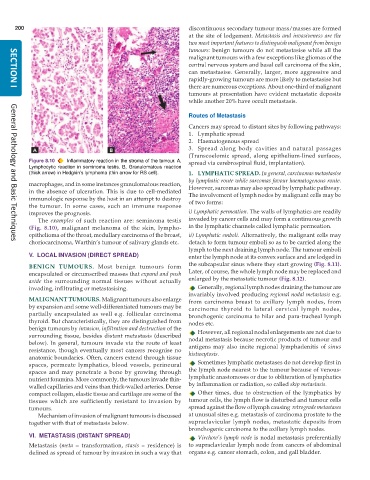
           Figure 8.10  Inflammatory reaction in the stroma of the tumour. A,  spread via cerebrospinal fluid, implantation).
           Lymphocytic reaction in seminoma testis. B, Granulomatous reaction
           (thick arrow) in Hodgkin’s lymphoma (thin arrow for RS cell).  1. LYMPHATIC SPREAD. In general, carcinomas metastasise
                                                               by lymphatic route while sarcomas favour haematogenous route.
           macrophages, and in some instances granulomatous reaction,  However, sarcomas may also spread by lymphatic pathway.
           in the absence of ulceration. This is due to cell-mediated  The involvement of lymph nodes by malignant cells may be
           immunologic response by the host in an attempt to destroy  of two forms:
           the tumour. In some cases, such an immune response
           improves the prognosis.                             i) Lymphatic permeation. The walls of lymphatics are readily
              The  examples  of such reaction are: seminoma testis  invaded by cancer cells and may form a continuous growth
           (Fig. 8.10), malignant melanoma of the skin, lympho-  in the lymphatic channels called lymphatic permeation.
           epithelioma of the throat, medullary carcinoma of the breast,  ii) Lymphatic emboli. Alternatively, the malignant cells may
           choriocarcinoma, Warthin’s tumour of salivary glands etc.  detach to form tumour emboli so as to be carried along the
     General Pathology and Basic Techniques
                                                               lymph to the next draining lymph node. The tumour emboli
           V.  LOCAL INVASION (DIRECT SPREAD)                  enter the lymph node at its convex surface and are lodged in
           BENIGN TUMOURS.  Most benign tumours form           the subcapsular sinus where they start growing (Fig. 8.11).
           encapsulated or circumscribed masses that expand and push  Later, of course, the whole lymph node may be replaced and
           aside  the surrounding normal tissues without actually  enlarged by the metastatic tumour (Fig. 8.12).
           invading, infiltrating or metastasising.               Generally, regional lymph nodes draining the tumour are
                                                               invariably involved producing regional nodal metastasis e.g.
           MALIGNANT TUMOURS. Malignant tumours also enlarge   from carcinoma breast to axillary lymph nodes, from
           by expansion and some well-differentiated tumours may be  carcinoma thyroid to lateral cervical lymph nodes,
           partially encapsulated as well e.g. follicular carcinoma  bronchogenic carcinoma to hilar and para-tracheal lymph
           thyroid. But characteristically, they are distinguished from  nodes etc.
           benign tumours by invasion, infiltration and destruction of the  However, all regional nodal enlargements are not due to
           surrounding tissue, besides distant metastasis (described  nodal metastasis because necrotic products of tumour and
           below). In general, tumours invade via the route of least
           resistance, though eventually most cancers recognise no  antigens may also incite regional lymphadenitis of sinus
           anatomic boundaries. Often, cancers extend through tissue  histiocytosis.
           spaces, permeate lymphatics, blood vessels, perineural  Sometimes lymphatic metastases do not develop first in
           spaces and may penetrate a bone by growing through  the lymph node nearest to the tumour because of venous-
           nutrient foramina. More commonly, the tumours invade thin-  lymphatic anastomoses or due to obliteration of lymphatics
           walled capillaries and veins than thick-walled arteries. Dense  by inflammation or radiation, so called skip metastasis.
           compact collagen, elastic tissue and cartilage are some of the  Other times, due to obstruction of the lymphatics by
           tissues which are sufficiently resistant to invasion by  tumour cells, the lymph flow is disturbed and tumour cells
           tumours.                                            spread against the flow of lymph causing  retrograde metastases
              Mechanism of invasion of malignant tumours is discussed  at unusual sites e.g. metastasis of carcinoma prostate to the
           together with that of metastasis below.             supraclavicular lymph nodes, metastatic deposits from
                                                               bronchogenic carcinoma to the axillary lymph nodes.
           VI.  METASTASIS (DISTANT SPREAD)                       Virchow’s lymph node is nodal metastasis preferentially
           Metastasis (meta  = transformation, stasis = residence) is  to supraclavicular lymph node from cancers of abdominal
           defined as spread of tumour by invasion in such a way that  organs e.g. cancer stomach, colon, and gall bladder.

