


Page 214 - Textbook of Pathology, 6th Edition
P. 214
198


     SECTION I


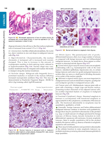
           Figure 8.5  Microscopic appearance of loss of nuclear polarity (B)
           contrasted with normal basal polarity in columnar epithelium (A). The
           basement membrane is intact in both.

           disproportionate to the cell size so that the nucleocytoplasmic
           ratio is increased from normal 1:5 to 1:1 (Fig. 8.6).
           iv) Anisonucleosis. Just like cellular pleomorphism, the nuclei  Figure 8.7  Normal and abnormal (atypical) mitotic figures.
           too, show variation in size and shape in malignant tumour
           cells (Fig. 8.6).                                   vii)  Mitotic figures.  The parenchymal cells of poorly-
                                                               differentiated tumours often show large number of mitoses
           v) Hyperchromatism.  Characteristically, the nuclear
           chromatin of malignant cell is increased and coarsely  as compared with benign tumours and well-differentiated
           clumped. This is due to increase in the amount of   malignant tumours. As stated above, these appear as either
           nucleoprotein resulting in dark-staining nuclei, referred to  normal or abnormal mitotic figures (Fig. 8.7):
           as hyperchromatism  (Fig. 8.6). Nuclear shape may vary,  Normal mitotic figures may be seen in some non-neoplastic
           nuclear membrane may be irregular and nuclear chromatin  proliferating cells (e.g. haematopoietic cells of the bone
           is clumped along the nuclear membrane.              marrow, intestinal epithelium, hepatocytes etc), in certain
                                                               benign tumours and some low grade malignant tumours; in
     General Pathology and Basic Techniques
           vi) Nucleolar changes. Malignant cells frequently have a  sections they are seen as a dark band of dividing chromatin
           prominent nucleolus or nucleoli in the nucleus reflecting  at two poles of the nuclear spindle.
           increased nucleoprotein synthesis (Fig. 8.6). This may be  Abnormal or atypical mitotic figures are more important in
           demonstrated as Nucleolar Organiser Region  (NOR) by  malignant tumours and are identified as tripolar, quadripolar
           silver (Ag) staining called AgNOR material.         and multipolar spindles in malignant tumour cells.

                                                               viii) Tumour giant cells. Multinucleate  tumour giant cells or
                                                               giant cells containing a single large and bizarre nucleus,
                                                               possessing nuclear characters of the adjacent tumour cells,
                                                               are another important feature of anaplasia in malignant
                                                               tumours (Fig. 8.8).
                                                               ix) Functional (Cytoplasmic) changes. Structural anaplasia in
                                                               tumours is accompanied with functional anaplasia as
                                                               appreciated from the cytoplasmic constituents of the tumour
                                                               cells. The functional abnormality in neoplasms may be
                                                               quantitative, qualitative, or both.
                                                                  Generally, benign tumours and better-differentiated
                                                               malignant tumours continue to function well qualitatively,
                                                               though there may be quantitative abnormality in the product
                                                               e.g. large or small amount of collagen produced by benign
                                                               tumours of fibrous tissue, keratin formation in well-
                                                               differentiated squamous cell carcinoma. In more anaplastic
                                                               tumours, there is usually quantitative fall in the product
                                                               made by the tumour cells e.g. absence of keratin in anaplastic
                                                               squamous cell carcinoma.
                                                                  There may be both qualitative and quantitative abnormality
                                                               of the cellular function in some anaplastic tumours e.g.
           Figure 8.6  Nuclear features of malignant cells in malignant  multiple myeloma producing abnormal immunoglobulin in
           melanoma—pleomorphism, anisonucleosis, increased N/C: ratio, nuclear
           hyperchromatism and prominent nucleoli.             large quantities.

