


Page 217 - Textbook of Pathology, 6th Edition
P. 217
201


                                                                                                                      CHAPTER 8


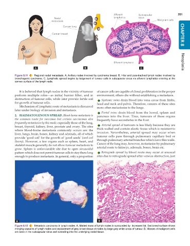
           Figure 8.11  Regional nodal metastasis. A, Axillary nodes involved by carcinoma breast. B, Hilar and para-tracheal lymph nodes involved by  Neoplasia
           bronchogenic carcinoma. C, Lymphatic spread begins by lodgement of tumour cells in subcapsular sinus via afferent lymphatics entering at the
           convex surface of the lymph node.


              It is believed that lymph nodes in the vicinity of tumour  of cancer cells are capable of clonal proliferation in the proper
           perform multiple roles—as initial barrier filter, and in  environment; others die without establishing a metastasis.
           destruction of tumour cells, while later provide fertile soil  Systemic veins drain blood into vena cavae from limbs,
           for growth of tumour cells.                         head and neck and pelvis. Therefore, cancers of these sites
              Mechanism of lymphatic route of metastasis is discussed  more often metastasise to the lungs.
           later under biology of invasion and metastasis.
                                                                  Portal veins drain blood from the bowel, spleen and
           2. HAEMATOGENOUS SPREAD. Blood-borne metastasis is  pancreas into the liver. Thus, tumours of these organs
           the common route for sarcomas but certain carcinomas also  frequently have secondaries in the liver.
           frequently metastasise by this mode, especially those of the lung,
           breast, thyroid, kidney, liver, prostate and ovary. The sites  Arterial spread of tumours is less likely because they are
           where blood-borne metastasis commonly occurs are: the  thick-walled and contain elastic tissue which is resistant to
           liver, lungs, brain, bones, kidney and adrenals, all of which  invasion. Nevertheless, arterial spread may occur when
           provide ‘good soil’ for the growth of ‘good seeds’ (seed-soil  tumour cells pass through pulmonary capillary bed or
           theory). However, a few organs such as spleen, heart, and  through pulmonary arterial branches which have thin walls.
           skeletal muscle generally do not allow tumour metastasis to  Cancer of the lung may, however, metastasise by pulmonary
           grow. Spleen is unfavourable site due to open sinusoidal  arterial route to kidneys, adrenals, bones, brain etc.
           pattern which does not permit tumour cells to stay there long  Retrograde spread by blood route may occur at unusual
           enough to produce metastasis. In general, only a proportion  sites due to retrograde spread after venous obstruction, just


           Figure 8.12  Metastatic carcinoma in lymph nodes. A, Matted mass of lymph nodes is surrounded by  increased fat. Sectioned surface shows
           merging capsules of lymph nodes and replacement of grey brown tissue of nodes by large grey white areas of tumour. B, Masses of malignant cells
           are seen in the subcapsular sinus and extending into the underlying nodal tissue.

