


Page 284 - Textbook of Pathology, 6th Edition
P. 284
268 different methods depending upon the purpose for which
they are intended:
i) Lateral vaginal smears (LVS) obtained by scraping the
upper third of the lateral walls of the vagina are ideal for
cytohormonal assessment.
ii) Vaginal ‘pool’ or ‘vault’ smears are obtained by scraping or
aspirating material from the posterior fornix of the vagina
and are recommended for detection of endometrial and
ovarian cancer.
SECTION I
iii) Cervical smears obtained by cotton swabs or Ayre’s spatula
from the portio of the cervix are ideal for detection of cervical
carcinoma.
iv) Combined (Fast) smears are a combination of vaginal pool
and cervical scrapings. They offer the advantages of both
and are recommended for routine population screening as
they allow detection of up to 97% of cervical cancers and
about 90% of endometrial cancers when properly prepared.
v) Triple (cervical-vaginal-endocervical or CVE) smears contain
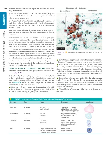
three distinct samples representing the ectocervix, vagina and Figure 11.1 Various types of epithelial cells seen in normal Pap
endocervix on three separate areas of the same slide. These smear.
smears are also recommended for routine screening as they
allow localisation of lesions but are difficult to prepare.
Lactation cells are parabasal cells with strongly acidophilic
vi) Endocervical and endometrial smears may also be prepared cytoplasm. These cells are seen so long as lactation persists.
by aspirating the contents of the endocervical canal and
endometrial cavity respectively. Endocervical cells appear either as single dispersed nuclei
due to degeneration, or as clusters of columnar cells giving
CELLS IN NORMAL COMBINED SMEARS. Normally, it honey-combed appearance. Nuclei of endocervical cells are
combined smear contains two types of cells: epithelial and vesicular, with fine granular chromatin and contain 1-2
others (Fig. 11.1): nucleoli, while the cytoplasm is slightly basophilic or
General Pathology and Basic Techniques
Epithelial cells. There are 4 types of squamous epithelial cells vacuolated.
in the cervical smear: superficial, intermediate, parabasal and Endometrial cells are seen up to 12th day of menstrual
basal cells. Morphological features of these cells are summed cycle. They are slightly smaller than endocervical cells,
up in Table 11.4. A few variants of morphological forms and appear as tight rounded clusters of overlapping cells with
other epithelial cells are as under: moderately dark oval nuclei and scanty basophilic,
Navicular cells are boat-shaped intermediate cells with vacuolated cytoplasm.
folded cell borders. These cells appear in latter half of the Trophoblastic cells are seen following abortion or after
menstrual cycle, during pregnancy and menopause. delivery.
TABLE 11.4: Squamous Epithelial Cells Found in Normal Combined (Fast) Smears.
Cell Type Size Nuclei Cytoplasm Morphology
Superficial 30-60 μm < 6 μm Polyhedral, thin, broad,
dark, pyknotic acidophilic or cyanophilic
with keratohyaline granules.
Intermediate 20-40 μm 6-9 μm Polyhedral or elongated,
vesicular thin, cyanophilic with
folded edges.
Parabasal 15-25 μm 6-11 μm Round to oval, thick,
vesicular well-defined, basophilic
with occasional small
vacuoles.
Basal 13-20 μm Large, (> one-half of cell Round to oval,
volume), hyperchromatic, deeply basophilic.
may have small nucleoli

