


Page 285 - Textbook of Pathology, 6th Edition
P. 285
Other cells. Besides epithelial cells, other cells in cervical TABLE 11.5: Representative Maturation Indices at Different 269
smears are leucocytes and Döderlein bacilli: Stages of Life.
Leucocytes in cervical smear include polymorphonuclear Stage Maturation Index* (%) Comment
neutrophils (in large numbers normally), lymphocytes
(isolated and entrapped in mucus normally), plasma cells Neonatal 0/90/10 As in pregnancy
(in chronic cervicitis), macrophages (normally in first 10 days Infancy 90/10/0 With infections
of the menstrual cycle) and multinucleate cells (in specific shows midzone CHAPTER 11
shift
inflammation).
Preovulatory 0/40/60 Shift-to-right
Döderlein bacilli (Bacillus vaginalis/Lactobacillus acidophilis),
which belong to the group of lactobacilli, are the predominant Post-ovulatory 0/70/30 Midzone shift
organisms of the normal vaginal flora. It is a slender, gram- Pregnancy 0/95/5 Midzone shift
positive, rod-like organism staining pale blue with the Postpartum 90/10/0 Shift-to-left
Papanicolaou technique. These organisms utilise the Menopausal 0/80/10 Estatrophy
glycogen contained in the cytoplasm of intermediate and (early)
parabasal cells resulting in their disintegration (cytolysis). Menopausal 95/5/0 Teleatrophy
They are most numerous in the luteal phase and during (late)
pregnancy. *MI = Parabasal/intermediate/superficial Basic Diagnostic Cytology
APPLICATIONS OF PAP SMEAR smears having sufficient number of evenly spread, well
preserved cells as evaluated microscopically.
Cytohormonal Evaluation
2. General categorisation: It includes categorising the smear
Assessment of hormonal status is best carried out from lateral in one of the three broad categories: within normal limits,
vaginal smears although vaginal ‘pool’ or fast smears may benign cellular changes, and epithelial cell abnormalities.
also be used. Ideally, at least 3 smears obtained on alternate
days should be scrutinised and cytologic indices determined 3. Descriptive diagnosis: Final aspect of the Bethesda
for each smear for accurate assessment. system includes detailed description of the benign cellular
Several indices are available for description of cyto- changes or epithelial cell abnormalities in the smear.
hormonal patterns. The most commonly used are as under: Based on it, the cellular changes in cervical smears are
i) Acidophilic index (AI): The relative proportion of cells described under 2 headings: non-neoplastic (or benign) and
containing acidophilic (pink) and basophilic (blue) cytoplasm neoplastic epithelial cell abnormalities.
are determined by AI.
ii) Pyknotic index (PI): The percentage of cells having small, NON-NEOPLASTIC (BENIGN) CELLULAR CHANGES:
dark, shrunken nuclei (less than 6 μm in size) is determined i) NON-SPECIFIC INFLAMMATORY CHANGES.
by PI. The PI is more reliable than AI as it is not influenced Inflammatory changes not associated with any specific
by many of the cytoplasmic artefacts e.g. acidophilia caused infection or identifiable infectious agent (i.e. non-specific
by drying of smears prior to fixation that affects the latter. inflammation) are commonly seen in smears of the female
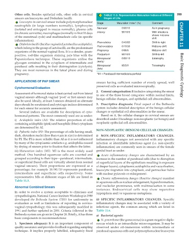
iii) Maturation index (MI): MI is the most widely used genital tract as under:
method. One hundred squamous cells are counted and Acute inflammatory changes are characterised by an
grouped according to their type—parabasal, intermediate, increase in the number of parabasal cells (due to disruption
or superficial (basal cells are virtually absent from normal of superficial layers of the epithelium resulting in exposure
vaginal smears). Their proportions are expressed as a of deeper layers), cytoplasmic acidophilia and vacuolisation,
percentage; for example 10/80/10 represents parabasal, leucocytic migration into cytoplasm, and perinuclear halos
intermediate and superficial cells respectively. Some with nuclear pyknosis or enlargement.
representative MIs at different stages of life are listed in Chronic inflammatory changes (Reactive changes) manifest
Table 11.5.
in squamous cells as nuclear enlargement, hyperchromatism,
Abnormal Combined Smears and nucleolar prominence, with multinucleation in some
instances. Endocervical cells may show reparative
In order to evolve a system acceptable to clinicians and hyperplasia and/or squamous metaplasia.
cytopathologists, National Cancer Institute Workshop in 1988
developed the Bethesda System (TBS) for uniformity in ii) SPECIFIC INFLAMMATORY CHANGES. Specific
evaluation as well as limitations of reporting in cervico- inflammatory changes may be associated with a variety of
vaginal cytopathology; this was subsequently modified in infectious agents, the common among which are listed in
1991 and further updated in 2001. Criteria followed in the Table 11.6.
Bethesda system are given in Chapter 26. Briefly, it has three a) Bacterial agents:
basic components in recommnedations:
N. gonorrhoeae (the gonococcus) is a gram-negative diplo-
1. Specimen adequacy: It is an important component of coccus which is an intracellular micro-organism. It may be
quality assurance and provides feedback regarding sampling observed under oil-immersion within intermediate or
technique. It implies properly labelled, adequately fixed parabasal squamous cells and polymorphonuclear leucocytes

