


Page 288 - Textbook of Pathology, 6th Edition
P. 288
272 Endocervical and endometrial adenocarcinoma, both of which
can be detected from Pap smears; cytomorphological features
allowing distinction between these two types of malignancies
are summed up in Table 11.7.
Cells from extrauterine cancers may also be present in Pap
smears, majority originating from the ovaries. Clues to the
extrauterine origin of cancer cells include: the absence of a
tumour diathesis, arrangement of cancer cells in glandular
and papillary configuration (accompanied by psammoma
SECTION I
bodies in some instances), and absence of dysplastic changes
in coexistent cervico-endometrial cells.
Automation in Cervical Cytology
Introduction of automated devices like PapNet for primary
screening is a major technologic achievement in recent
times. Automation offers routine pre-screening of hundreds
of Pap smears, decreasing the workload of cytopathologists
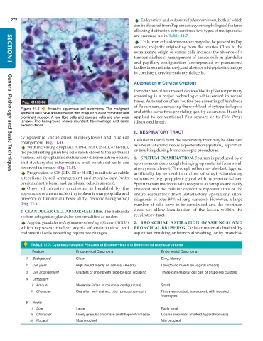
Figure 11.6 Invasive squamous cell carcinoma. The malignant and at the same time providing quality assurance. It can be
epithelial cells have anisonucleosis with irregular nuclear chromatin with
prominent nucleoli. A few fibre cells and caudate cells are also seen applied to conventional Pap smears or to Thin-Preps
(arrow). The background shows abundant haemorrhage and some (discussed later).
necrotic debris.
II. RESPIRATORY TRACT
cytoplasmic vacuolation (koilocytosis) and nuclear Cellular material from the respiratory tract may be obtained
enlargement (Fig. 11.4).
With increasing dysplasia (CIN-II and CIN-III, or H-SIL), as a result of spontaneous expectoration (sputum), aspiration
the proliferating primitive cells reach closer to the epithelial or brushing during bronchoscopic procedures.
surface, less cytoplasmic maturation/differentiation occurs, 1. SPUTUM EXAMINATION. Sputum is produced by a
and dyskaryotic intermediate and parabasal cells are spontaneous deep cough bringing up material from small
observed in smears (Fig. 11.5). airways and alveoli. The cough reflex may also be triggered
Progression to CIS (CIN-III or H-SIL) manifests as subtle artificially by aerosol-inhalation of cough-stimulating
General Pathology and Basic Techniques
alterations in cell arrangement and morphology (with substances (e.g. propylene glycol with hypertonic saline).
predominantly basal and parabasal cells in smears). Sputum examination is advantageous as samples are easily
Onset of invasive carcinoma is heralded by the obtained and the cellular content is representative of the
appearance of macronucleoli, cytoplasmic orangeophilia and entire respiratory tract (satisfactory specimens allow
presence of tumour diathesis (dirty, necrotic background) diagnosis of over 80% of lung cancers). However, a large
(Fig. 11.6). number of cells have to be scrutinised and the specimen
2. GLANDULAR CELL ABNORMALITIES. The Bethesda does not allow localisation of the lesion within the
system categorises glandular abnormalities as under: respiratory tract.
Atypical glandular cells of undetermined significance (AGUS) 2. BRONCHIAL ASPIRATION (WASHINGS) AND
which represent nuclear atypia of endocervical and BRONCHIAL BRUSHING. Cellular material obtained by
endometrial cells exceeding reparative changes. aspiration brushing or bronchial washing, or by bronchio-
TABLE 11.7: Cytomorphological Features of Endocervical and Endometrial Adenocarcinoma.
Feature Endocervical Carcinoma Endometrial Carcinoma
1. Background Clean Dirty, bloody
2. Cell yield High (found mainly on cervical smears) Low (found mainly on vaginal smears)
3. Cell arrangement Clusters or sheets with ‘side-by-side’ grouping Three-dimensional ‘cell ball’ or grape-like clusters
4. Cytoplasm
i) Amount Moderate (often in columnar configuration) Small
ii) Character Granular, well-stained, often producing mucin Finely vacuolated, translucent, with ingested
leucocytes
5. Nuclei
i) Size Large Fairly small
ii) Character Finely granular chromatin (mild hyperchromasia) Coarse chromatin (marked hyperchromasia)
iii) Nucleoli Macronucleoli Micronucleoli

