


Page 287 - Textbook of Pathology, 6th Edition
P. 287
271


                                                                                                                      CHAPTER 11


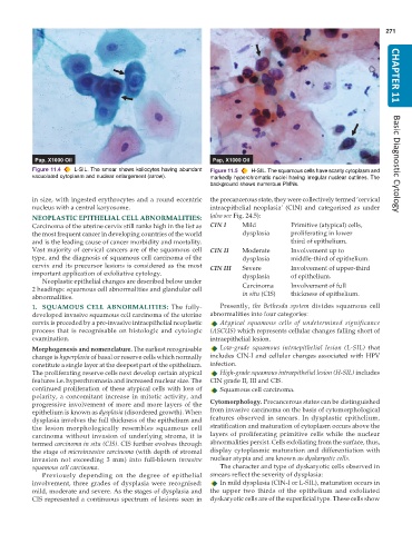
           Figure 11.4  L-SIL. The smear shows koilocytes having abundant  Figure 11.5  H-SIL. The squamous cells have scanty cytoplasm and  Basic Diagnostic Cytology
           vacuolated cytoplasm and nuclear enlargement (arrow).  markedly hyperchromatic nuclei having irregular nuclear outlines. The
                                                               background shows numerous PMNs.
           in size, with ingested erythrocytes and a round eccentric  the precancerous state, they were collectively termed ‘cervical
           nucleus with a central karyosome.                   intraepithelial neoplasia’ (CIN) and categorised as under
           NEOPLASTIC EPITHELIAL CELL ABNORMALITIES:           (also see Fig. 24.5):
           Carcinoma of the uterine cervix still ranks high in the list as  CIN I  Mild  Primitive (atypical) cells,
           the most frequent cancer in developing countries of the world  dysplasia    proliferating in lower
           and is the leading cause of cancer morbidity and mortality.                 third of epithelium.
           Vast majority of cervical cancers are of the squamous cell  CIN II  Moderate  Involvement up to
           type, and the diagnosis of squamous cell carcinoma of the     dysplasia     middle-third of epithelium.
           cervix and its precursor lesions is considered as the most  CIN III  Severe  Involvement of upper-third
           important application of exfoliative cytology.                dysplasia     of epithelium.
              Neoplastic epithelial changes are described below under
           2 headings: squamous cell abnormalities and glandular cell    Carcinoma     Involvement of full
           abnormalities.                                                in situ (CIS)  thickness of epithelium.
           1. SQUAMOUS CELL ABNORMALITIES:  The fully-            Presently, the  Bethesda system divides squamous cell
           developed invasive squamous cell carcinoma of the uterine  abnormalities into four categories:
           cervix is preceded by a pre-invasive intraepithelial neoplastic  Atypical squamous cells of undetermined significance
           process that is recognisable on histologic and cytologic  (ASCUS) which represents cellular changes falling short of
           examination.                                        intraepithelial lesion.
           Morphogenesis and nomenclature. The earliest recognisable  Low-grade squamous intraepithelial lesion (L-SIL)  that
           change is hyperplasia of basal or reserve cells which normally  includes CIN-I and cellular changes associated with HPV
           constitute a single layer at the deepest part of the epithelium.  infection.
           The proliferating reserve cells next develop certain atypical  High-grade squamous intraepithelial lesion (H-SIL) includes
           features i.e. hyperchromasia and increased nuclear size. The  CIN grade II, III and CIS.
           continued proliferation of these atypical cells with loss of  Squamous cell carcinoma.
           polarity, a concomitant increase in mitotic activity, and
           progressive involvement of more and more layers of the  Cytomorphology. Precancerous states can be distinguished
           epithelium is known as dysplasia (disordered growth). When  from invasive carcinoma on the basis of cytomorphological
           dysplasia involves the full thickness of the epithelium and  features observed in smears. In dysplastic epithelium,
           the lesion morphologically resembles squamous cell  stratification and maturation of cytoplasm occurs above the
           carcinoma without invasion of underlying stroma, it is  layers of proliferating primitive cells while the nuclear
           termed carcinoma in situ (CIS). CIS further evolves through  abnormalities persist. Cells exfoliating from the surface, thus,
           the stage of microinvasive carcinoma (with depth of stromal  display cytoplasmic maturation and differentiation with
           invasion not exceeding 3 mm) into full-blown  invasive  nuclear atypia and are known as dyskaryotic cells.
           squamous cell carcinoma.                               The character and type of dyskaryotic cells observed in
              Previously depending on the degree of epithelial  smears reflect the severity of dysplasia:
           involvement, three grades of dysplasia were recognised:  In mild dysplasia (CIN-I or L-SIL), maturation occurs in
           mild, moderate and severe. As the stages of dysplasia and  the upper two thirds of the epithelium and exfoliated
           CIS represented a continuous spectrum of lesions seen in  dyskaryotic cells are of the superficial type. These cells show

