


Page 286 - Textbook of Pathology, 6th Edition
P. 286
270 aggregates of rounded to oval squamous cells with blunted
TABLE 11.6: Common Infections Detectable on Pap Smears.
cell margins, peripheral cytoplasmic condensation and
BACTERIAL AGENTS crisply outlined perinuclear halos, with dysplastic or
Neisseria gonorrhoeae degenerative changes of nuclei; these changes are collectively
Gardnerella vaginalis
Mycobacterium tuberculosis termed ‘koilocytotic changes.’ (see Fig. 11.4). HPV can also
VIRAL AGENTS be identified by Southern blot, PCR, in situ hybridization and
Herpes simplex virus (HSV) by hybrid capture.
Human papillomavirus (HPV) Genital infection by herpes simplex virus (HSV) commonly
FUNGAL AGENTS affects the squamous epithelium of the vulva, vagina and
SECTION I
Candida albicans cervix, but may ascend to involve endocervical and
Torulopsis glabrata endometrial epithelium. Infected cells show enlarged nuclei
PARASITIC AGENTS with watery, ‘ground-glass’ chromatin, multinucleation with
Trichomonas vaginalis internuclear moulding, and intranuclear acidophilic
Entamoeba histolytica
inclusions.
c) Fungal agents:
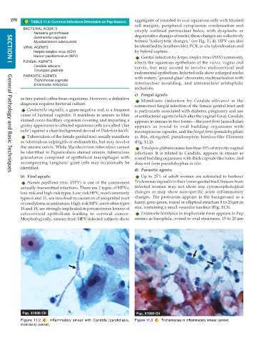
as tiny paired coffee-bean organisms. However, a definitive Moniliasis (infection by Candida albicans) is the
diagnosis requires bacterial culture.
commonest fungal infection of the female genital tract and
Gardnerella vaginalis, a gram-negative rod, is a frequent is particularly associated with diabetes, pregnancy and use
cause of bacterial vaginitis. It manifests in smears as blue of antibacterial agents (which alter the vaginal flora). Candida
stained cocco-bacillary organism covering and imparting a appears in smears in two forms—the yeast form (unicellular)
grainy appearance to affected squamous cells (so called ‘clue appears as round to oval budding organisms with
cells’) against a clean background devoid of Döderlein bacilli. inconspicuous capsules, and the fungal form (pseudohyphae)
Tuberculosis of the female genital tract usually manifests as thin, elongated, pseudoseptate, bamboo-like filaments
as tuberculous salpingitis or endometritis, but may involve (Fig. 11.2).
the uterine cervix. While Mycobacterium tuberculosis cannot Torulopsis glabrata causes less than 10% of mycotic vaginal
be identified in Papanicolaou stained smears, tuberculous infections. It is related to Candida, appears in smears as
granulomas composed of epithelioid macrophages with round budding organisms with thick capsule-like halos, and
accompanying Langhans’ giant cells may occasionally be does not form pseudohyphae in vivo.
identified.
d) Parasitic agents:
General Pathology and Basic Techniques
b) Viral agents: Up to 25% of adult women are estimated to harbour
Human papilloma virus (HPV) is one of the commonest Trichomonas vaginalis in their lower genital tract. Smears from
sexually-transmitted infections. There are 2 types of HPVs: infected women may not show any cytomorphological
low-risk and high-risk types. Low-risk HPV, most commonly changes or may show non-specific acute inflammatory
types 6 and 11, are involved in causation of anogenital wart changes. The protozoan appears in the background as a
or condyloma acuminatum. High-risk HPV, most often types fuzzy, grey-green, round or elliptical structure 8 to 20 μm in
16 and 18, are strongly implicated in precancerous lesions of size, containing a small vesicular nucleus (Fig. 11.3).
ectocervical epithelium leading to cervical cancer. Entamoeba histolytica in trophozoite form appears in Pap
Morphologically, smears from HPV-infected subjects show smears as basophilic, round to oval structures, 15 to 20 μm
Figure 11.2 Inflammatory smear with Candida (candidiasis, Figure 11.3 Trichomonas in inflammatory smear (arrow).
moniliasis) (arrow).

