


Page 290 - Textbook of Pathology, 6th Edition
P. 290
274                                                      ii) Correct cytological processing and technique, since the
                                                               cell yield of CSF is generally low and faulty technique may
                                                               result in loss of cellular material or poor morphology.
                                                               Normal CSF. CSF is an ultrafiltrate of plasma with a total
                                                               volume of about 150 ml (in the adult). Cell content is low
                                                               (0-4/μl) comprising mainly lymphocytes or monocytes.
                                                               CSF in non-neoplastic diseases. Changes in CSF in various
                                                               non-neoplastic diseases are discussed in Chapter 30 (see
                                                                Table 30.1).
     SECTION I
                                                               CSF in neoplastic disease. Neoplasms that are in direct
                                                               contact with the CSF are most likely to shed cells which are
                                                               recoverable for scrutiny. Metastatic cancers (leukaemia,
                                                               lymphoma and adenocarcinoma), medulloblastomas, and
                                                               ependymomas shed the greatest amount of material. Though
                                                               meningioma occurs in the subarachnoid space, it rarely
                                                               exfoliates sufficient material for diagnosis. Low-grade
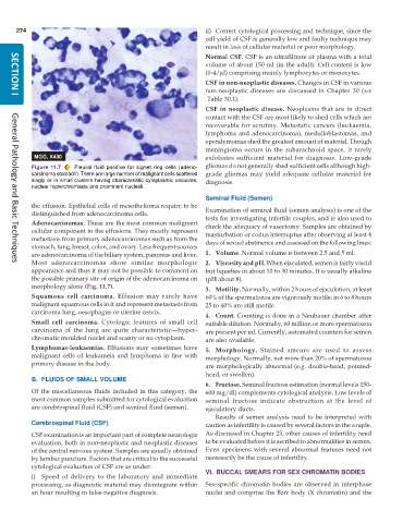
           Figure 11.7  Pleural fluid positive for signet ring cells (adeno-  gliomas do not generally shed sufficient cells although high-
           carcinoma stomach). There are large number of malignant cells scattered  grade gliomas may yield adequate cellular material for
           singly or in small clusters having characteristic cytoplasmic vacuoles,  diagnosis.
           nuclear hyperchromasia and prominent nucleoli.
                                                               Seminal Fluid (Semen)
           the effusion. Epithelial cells of mesothelioma require to be  Examination of seminal fluid (semen analysis) is one of the
           distinguished from adenocarcinoma cells.            tests for investigating infertile couples, and is also used to
           Adenocarcinomas. These are the most common malignant  check the adequacy of vasectomy. Samples are obtained by
           cellular component in the effusions. They mostly represent  masturbation or coitus interruptus after observing at least 4
           metastasis from primary adenocarcinomas such as from the  days of sexual abstinence and assessed on the following lines:
           stomach, lung, breast, colon, and ovary. Less frequent sources
           are adenocarcinoma of the biliary system, pancreas and liver.  1. Volume. Normal volume is between 2.5 and 5 ml.
           Most adenocarcinomas show similar morphologic       2. Viscosity and pH. When ejaculated, semen is fairly viscid
     General Pathology and Basic Techniques
           appearance and thus it may not be possible to comment on  but liquefies in about 10 to 30 minutes. It is usually alkaline
           the possible primary site of origin of the adenocarcinoma on  (pH about 8).
           morphology alone (Fig. 11.7).                       3. Motility. Normally, within 2 hours of ejaculation, at least
           Squamous cell carcinoma. Effusion may rarely have   60% of the spermatozoa are vigorously motile; in 6 to 8 hours
           malignant squamous cells in it and represent metastasis from  25 to 40% are still motile.
           carcinoma lung, oesophagus or uterine cervix.
                                                               4. Count. Counting is done in a Neubauer chamber after
           Small cell carcinoma. Cytologic features of small cell  suitable dilution. Normally, 60 million or more spermatozoa
           carcinoma of the lung are quite characteristic—hyper-  are present per ml. Currently, automated counters for semen
           chromatic moulded nuclei and scanty or no cytoplasm.  are also available.
           Lymphomas-leukaemias. Effusions may sometimes have  5. Morphology. Stained smears are used to assess
           malignant cells of leukameia and lymphoma in line with  morphology. Normally, not more than 20% of spermatozoa
           primary disease in the body.                        are morphologically abnormal (e.g. double-head, pointed-
                                                               head, or swollen).
           B. FLUIDS OF SMALL VOLUME
                                                               6. Fructose. Seminal fructose estimation (normal levels 150-
           Of the miscellaneous fluids included in this category, the  600 mg/dl) complements cytological analysis. Low levels of
           most common samples submitted for cytological evaluation  seminal fructose indicate obstruction at the level of
           are cerebrospinal fluid (CSF) and seminal fluid (semen).  ejaculatory ducts.
                                                                  Results of semen analysis need to be interpreted with
           Cerebrospinal Fluid (CSF)                           caution as infertility is caused by several factors in the couple.

           CSF examination is an important part of complete neurologic  As discussed in Chapter 23, other causes of infertility need
           evaluation, both in non-neoplastic and neoplastic diseases  to be evaluated before it is ascribed to abnormalities in semen.
           of the central nervous system. Samples are usually obtained  Even specimens with several abnormal features need not
           by lumbar puncture. Factors that are critical to the successful  necessarily be the cause of infertility.
           cytological evaluation of CSF are as under:
                                                               VI. BUCCAL SMEARS FOR SEX CHROMATIN BODIES
           i) Speed of delivery to the laboratory and immediate
           processing, as diagnostic material may disintegrate within  Sex-specific chromatin bodies are observed in interphase
           an hour resulting in false-negative diagnosis.      nuclei and comprise the Barr body (X chromatin) and the

