


Page 291 - Textbook of Pathology, 6th Edition
P. 291
fluorescent or F body (Y chromatin). The number of Barr                                                  275
           bodies observed in interphase nuclei is one less than the
           number of X chromosome (n–1), whereas the number of F
           bodies observed is equal to the number of Y chromosomes
           present (n). In effect, the Barr body is specific for females
           and the F body for males (page 257).                                                                       CHAPTER 11
           1. DEMONSTRATION OF BARR BODY (X CHROMA-
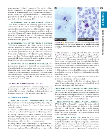
           TIN). In buccal smears, the Barr body appears as a plano-
           convex mass about 1 μm in diameter applied to the inner
           surface of the nuclear membrane. The interphase nuclei of
           one hundred intermediate squamous epithelial cells are
           scrutinised. In normal females, Barr bodies are present in at
           least 20% of nuclei (Fig. 11.8, A). Males have a Barr body
           count of less than 2%. Vaginal smears may also be used for
           Barr body counts (page 257).
           2. DEMONSTRATION OF THE F BODY (Y CHROMA-           Figure 11.8  Sex chromatin. A, Buccal smear showing condensation  Basic Diagnostic Cytology
           TIN).  Demonstration of the F body requires fluorescent  of chromatin under the nuclear membrane. B, Blood film showing
                                                               presence of drumstick appendage attached to a nuclear lobe of the
           staining in contrast to the Barr body which may be observed  neutrophil.
           on routine Papanicolaou-stained smears. On staining buccal
           smears with quinacrine mustard, the intensely fluorescent F
           body is observed in about 60% of interphase nuclei in males  iv) The ectocervix is sampled with the  Ayre’s spatula
                                                               (Fig. 11.9). The longer limb of the spatula is fitted into the
           and in less than 8% of nuclei in females. The F body is also  external os and the spatula rotated through 360° to sample
           demonstrable in the nuclei of lymphocytes in a peripheral  the entire cervix. The scraped material on the spatula is then
           blood film stained with quinacrine mustard.
                                                               placed on the drop aspirated from the vaginal pool and the
           3. COUNTING OF DRUMSTICK APPENDAGE.  The            smear prepared with the spatula itself or with the tip of the
           presence and frequency of drumstick appendages attached  gloved finger (cotton swabs may be used instead of the Ayre’s
           to nuclei of polymorphonuclear leucocytes on a peripheral  spatula, although the quality of smears is not as good).
           blood film may also be used for determining sex. At least  v) Thin uniform smears should be prepared and the slide
           500 neutrophilic leucocytes are scrutinised in a Romanowsky-  immediately immersed in fixative to avoid artefacts in cells
           stained blood film. Genetic females show drumsticks in 3-  caused by drying.
           6% of neutrophils (Fig. 11.8, B). In males, the frequency of  vi) Smears should be  transported  to the laboratory in the
           drumsticks is less than 0.3%.                       fixative in a Coplin’s jar (discussed later).
                                                                  Slides should ideally be labelled by using a diamond
           TECHNIQUES IN EXFOLIATIVE CYTOLOGY                  pencil. If labels of sticking plaster are used, the labels must
                                                               not come into contact with the fixative.
           A brief resume of methods for collection of specimens and
           processing of samples for exfoliative cytodiagnosis is  2. LIQUID-BASED CYTOLOGY PREPARATIONS (THIN
           outlined below under 4 headings: collection, fixation and  PREPS).  This is a special technique for preparation of
           fixatives, processing in the laboratory, and staining.  gynaecologic and non-gynaecologic samples which provides
                                                               uniform monolayered dispersion of cells on smears, without
                                                               overlapping or clump formation. It is a pre-requisite for
           A. Collection of Samples
                                                               quantitative analysis and automated devices.
           1. PREPARATION OF COMBINED (FAST) SMEARS.
           Smears should not be taken during menstrual bleeding. The  3. PREPARATION OF LATERAL VAGINAL SMEARS
           patient should not douche for at least 24 hours before the  (LVS). The LVS is obtained by scraping the lateral walls of
           smear is obtained.                                  the upper third of the vagina (at the level of the cervical
                                                               external os) with the flat surface of a wooden tongue
           i) Smears are obtained under direct vision after introducing  depressor and smearing the material directly onto labelled
           a Cusco’s speculum with the patient in lithotomy position.  slides. The patient is prepared and positioned as described
           ii) Ideally, lubricants and medical jellies should not be used  for the combined (fast) smear.
           for introducing the speculum since use of lubricants results  4. COLLECTION OF SPUTUM.  Fresh, unfixed, early
           in contamination of the smear. If required, the speculum may  morning specimens resulting from overnight accumulation
           be moistened with a few drops of normal saline.
                                                               of secretions are best for diagnostic purposes. A minimum
           iii) The posterior fornix of the vagina is aspirated with a  of at least three specimens collected on three successive days
           blunt-ended glass pipette fitted with a rubber bulb. A drop of  should be examined. The patient is instructed to cough
           the aspirate is placed at the unlabelled end of a glass slide.  deeply on waking up and to expectorate all sputum into a

