


Page 295 - Textbook of Pathology, 6th Edition
P. 295
viii) Following withdrawal of the needle from the lesion,  279
                                                               pressure is applied to the site of puncture by the assistant or
                                                               patient himself for 2 to 3 minutes in order to arrest bleeding
                                                               and prevent haematoma formation.
                                                               ix) Aspirated material is recovered by detaching the needle
                                                               from the syringe and filling the syringe with air; the syringe  CHAPTER 11
                                                               and needle are then reconnected and the aspirate expressed
                                                               onto one end of a glass slide.

                                                               Preparation of Smears

                                                               Preparation of smears is crucial to the success of FNAC.
                                                               Poorly-prepared smears with distorted cellular morphology
                                                               will frustrate the best efforts of the most competent
                                                               cytopathologist, and often result in errors of interpretation
                                                               or in failure to arrive at any specific diagnosis. The procedure
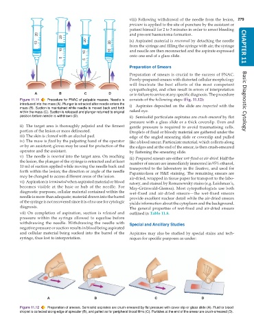
           Figure 11.11  Procedure for FNAC of palpable masses. Needle is  consists of the following steps (Fig. 11.12):  Basic Diagnostic Cytology
           introduced into the mass (A). Plunger is retracted after needle enters the  i) Aspirates deposited on the slide are inspected with the
           mass (B). Suction is maintained while needle is moved back and forth
           within the mass (C). Suction is released and plunger returned to original  naked eye.
           position before needle is withdrawn (D).            ii) Semisolid particulate aspirates are crush-smeared by flat
                                                               pressure with a glass slide or a thick coverslip. Even and
           ii) The target area is thoroughly  palpated and the firmest  gentle pressure is required to avoid traumatising cells.
           portion of the lesion or mass delineated.           Droplets of fluid or bloody material are gathered under the
           iii) The skin is cleaned with an alcohol pad.       edge of the angled smearing slide or coverslip and pulled
           iv) The mass is fixed by the palpating hand of the operator  like a blood smear. Particulate material, which collects along
           or by an assistant; gloves may be used for protection of the  the edges and at the end of the smear, is then crush-smeared
           operator and the assistant.                         by flattening the smearing slide.
           v) The needle is inserted into the target area. On reaching  iii) Prepared smears are either wet-fixed or air-dried. Half the
           the lesion, the plunger of the syringe is retracted and at least  number of smears are immediately immersed in 95% ethanol,
           10 ml of suction applied while moving the needle back and  transported to the laboratory in the fixative, and used for
           forth within the lesion; the direction or angle of the needle  Papanicolaou or H&E staining. The remaining smears are
           may be changed to access different areas of the lesion.  air-dried, wrapped in tissue paper for transport to the labo-
           vi) Aspiration is terminated when aspirated material or blood  ratory, and stained by Romanowsky stains (e.g. Leishman’s,
           becomes visible at the base or hub of the needle. For  May-Grünwald-Giemsa). Most cytopathologists use both
           diagnostic purposes, cellular material contained within the  wet-fixed and air-dried smears—the wet-fixed smears
           needle is more than adequate; material drawn into the barrel  provide excellent nuclear detail while the air-dried smears
           of the syringe is not recovered since it is of no use for cytologic  yields information about the cytoplasm and the background.
           diagnosis.                                          The general properties of wet-fixed and air-dried smears
           vii) On completion of aspiration, suction is  released and  outlined in Table 11.8.
           pressure within the syringe allowed to equalise before
           withdrawing the needle. Withdrawing the needle with  Special and Ancillary Studies
           negative pressure or suction results in blood being aspirated
           and cellular material being sucked into the barrel of the  Aspirates may also be studied by special stains and tech-
           syringe, thus lost to interpretation.               niques for specific purposes as under:


           Figure 11.12  Preparation of smears. Semisolid aspirates are crush-smeared by flat pressure with cover slip or glass slide (A). Fluid or blood
           droplet is collected along edge of spreader (B), and pulled as for peripheral blood films (C). Particles at the end of the smear are crush-smeared (D).

