


Page 298 - Textbook of Pathology, 6th Edition
P. 298
282


     SECTION I


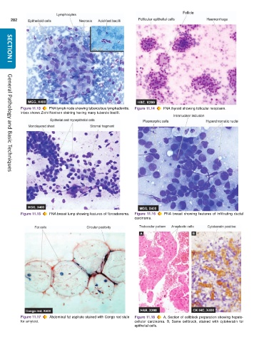
           Figure 11.13  FNA lymph node showing tuberculous lymphadenitis.  Figure 11.14  FNA thyroid showing follicular neoplasm.
           Inbox shows Ziehl-Neelsen staining having many tubercle bacilli.


     General Pathology and Basic Techniques


           Figure 11.15  FNA breast lump showing features of fibroadenoma.  Figure 11.16  FNA breast showing features of infiltrating ductal
                                                               carcinoma.


           Figure 11.17  Abdominal fat aspirate stained with Congo red stain  Figure 11.18  A, Section of cellblock preparation showing hepato-
           for amyloid.                                        cellular carcinoma. B, Same cellblock, stained with cytokeratin for
                                                               epithelial cells.

