


Page 304 - Textbook of Pathology, 6th Edition
P. 304
288


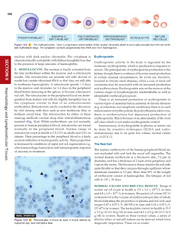
           Figure 12.5  The erythroid series. There is progressive condensation of the nuclear chromatin which is eventually extruded from the cell at the
           late erythroblast stage. The cytoplasm contains progressively less RNA and more haemoglobin.


           nucleus with dark nuclear chromatin. The cytoplasm is  Erythropoietin
           characteristically acidophilic with diffuse basophilic hue due
           to the presence of large amounts of haemoglobin.    Erythropoietic activity in the body is regulated by the
                                                               hormone, erythropoietin, which is produced in response to
           5. RETICULOCYTE. The nucleus is finally extruded from  anoxia. The principal site of erythropoietin production is the
           the late erythroblast within the marrow and a reticulocyte  kidney though there is evidence of its extra-renal production
           results. The reticulocytes are juvenile red cells devoid of  in certain unusual circumstances. Its levels are, therefore,
           nuclei but contain ribosomal RNA so that they are still able  lowered in chronic renal diseases, while a case of renal cell
     SECTION II
           to synthesise haemoglobin. A reticulocyte spends 1-2 days  carcinoma may be associated with its increased production
           in the marrow and circulates for 1-2 days in the peripheral  and erythrocytosis. Erythropoietin acts on the marrow at the
           blood before maturing in the spleen, to become a biconcave  various stages of morphologically unidentifiable as well as
           red cell. The reticulocytes in the peripheral blood are distin-  identifiable erythroid precursors.
           guished from mature red cells by slightly basophilic hue in  There is an increased production of erythropoietin in
           the cytoplasm similar to that of an orthochromatic  various types of anaemias but in anaemia of chronic diseases
           erythroblast. Reticulocytes can be counted in the laboratory  (e.g. in infections and neoplastic conditions) there is no such
           by vital staining with dyes such as new methylene blue or  enhancement of erythropoietin. In polycythaemia rubra vera,
           brilliant cresyl blue. The reticulocytes by either of these  there is erythrocytosis but depressed production of
           staining methods contain deep blue reticulofilamentous  erythropoietin. This is because of an abnormality of the stem
           material (Fig. 12.6). While erythroblasts are not normally  cell class which is not under erythropoietin control.
           present in human peripheral blood, reticulocytes are found  Immunoassay of erythropoietin in plasma or serum can
           normally in the peripheral blood. Normal range of   be done by sensitive techniques (ELISA and radio-
           reticulocyte count in health is 0.5-2.5% in adults and 2-6% in  immunoassay) due to its quite low values; normal values
           infants. Their percentage in the peripheral blood is a fairly  are 10-25 U/L.
           accurate reflection of erythropoietic activity. Their proportion
           is increased in conditions of rapid red cell regeneration e.g.  The Red Cell
           after haemorrhage, haemolysis and haematopoietic response  The mature erythrocytes of the human peripheral blood are
           of anaemia to treatment.
                                                               non-nucleated cells and lack the usual cell organelles. The
                                                               normal human erythrocyte is a biconcave disc, 7.2 μm in
                                                               diameter, and has a thickness of 2.4 μm at the periphery and
     Haematology and Lymphoreticular Tissues
                                                               1 μm in the centre. The biconcave shape renders the red cells
                                                               quite flexible so that they can pass through capillaries whose
                                                               minimum diameter is 3.5 μm. More than 90% of the weight
                                                               of erythrocyte consist of haemoglobin. The lifespan of red
                                                               cells is 120 + 30 days.

                                                               NORMAL VALUES AND RED CELL INDICES.  Range of
                                                                                                       12
                                                               normal red cell count in health is 5.5 ± 1.0 × 10 /L in men
                                                                             12
                                                               and 4.8 ± 1.0 × 10 /L in women. The packed cell volume (PCV)
                                                               or haematocrit is the volume of erythrocytes per litre of whole
                                                               blood indicating the proportion of plasma and red cells and
                                                               ranges 0.47 ± 0.07 L/L (40-54%) in men and 0.42 ± 0.05 L/L
                                                               (37-47%) in women. The haemoglobin content in health is 15.5
                                                               ± 2.5 g/dl (13-18 g/dl) in men and 14.0 ± 2.5 g/dl (11.5-16.5
                                                               g/dl) in women. Based on these normal values, a series of
           Figure 12.6  Reticulocytes in blood as seen in blood stained by  absolute values or red cell indices can be derived which have
           supravital dye, new methylene blue.                 diagnostic importance. These are as under:

