


Page 309 - Textbook of Pathology, 6th Edition
P. 309
293


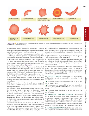
           Figure 12.10  Some of the common morphologic abnormalities of red cells (The serial numbers in the illustrations correspond to the order in  CHAPTER 12
           which they are described in the text).

           Pappenheimer bodies which stain positively). Classical  vii)  Acanthocytosis is the presence of coarsely crenated red
           punctate basophilia is seen in aplastic anaemia, thalassaemia,  cells. Acanthocytes are found in large number in blood film
           myelodysplasia, infections and lead poisoning.      made from splenectomised subjects, and in chronic liver
           iv) Howell-Jolly bodies are purple nuclear remnants, usually  disease.
           found singly, and are larger than basophilic stippling. They  viii) Burr cells are cell fragments having one or more spines.
           are present in megaloblastic anaemia and after splenectomy.  They are particularly found in uraemia.
           5. Miscellaneous changes. In addition to the morphologic  ix) Stomatocytosis is the presence of stomatocytes which have
           changes of red cells described above, several other abnormal  central area having slit-like or mouth-like appearance. They
           red cells may be found in different haematological disorders.  are found in hereditary stomatocytosis, or may be seen in
           Some of these are as follows (Fig. 12.10):          chronic alcoholism.
           i) Spherocytosis is characterised by presence of spheroidal  x) Ovalocytosis or elliptocytosis is the oval or elliptical shape
           rather than biconcave disc-shaped red cells. Spherocytes are  of red cells. Their highest proportion (79%) is seen in
           seen in hereditary spherocytosis, autoimmune haemolytic  hereditary ovalocytosis and elliptocytosis; other conditions
           anaemia and in ABO haemolytic disease of the newborn.  showing such abnormal shapes of red cells are megaloblastic  Introduction to Haematopoietic System and Disorders of Erythroid Series
           ii) Schistocytosis is identified by fragmentation of erythro-  anaemia and hypochromic anaemia.
           cytes. Schistocytes are found in thalassaemia, hereditary  C. RED CELL INDICES.  An alternative method to diagnose
           elliptocytosis, megaloblastic anaemia, iron deficiency  and detect the severity of anaemia is by measuring the red
           anaemia, microangiopathic haemolytic anaemia and in  cell indices:
           severe burns.                                          In iron deficiency and thalassaemia, MCV, MCH and
           iii) Irregularly contracted red cells are found in drug and  MCHC are reduced.
           chemical induced haemolytic anaemia and in unstable    In anaemia due to acute blood loss and haemolytic
           haemoglobinopathies.                                anaemias, MCV, MCH and MCHC are all within normal
           iv) Leptocytosis is the presence of unusually thin red cells.  limits.
           Leptocytes are seen in severe iron deficiency and      In megaloblastic anaemias, MCV is raised above the
           thalassaemia. Target cell is a form of leptocyte in which there  normal range.
           is central round stained area and a peripheral rim of
           haemoglobin. Target cells are found in iron deficiency,  D. LEUCOCYTE AND PLATELET COUNT.  Measurement
           thalassaemia, chronic liver disease, and after splenectomy.  of leucocyte and platelet count helps to distinguish pure
           v) Sickle cells or drepanocytes are sickle-shaped red cells found  anaemia from pancytopenia in which red cells, granulocytes
                                                               and platelets are all reduced. In anaemias due to haemolysis
           in sickle cell disease.                             or haemorrhage, the neutrophil count and platelet counts
           vi) Crenated red cells are the erythrocytes which develop  are often elevated. In infections and leukaemias, the leucocyte
           numerous projections from the surface. They are present in  counts are high and immature leucocytes appear in the blood.
           blood films due to alkaline pH, presence of traces of fatty
           substances on the slides and in cases where the film is made  E. RETICULOCYTE COUNT. Reticulocyte count (normal
           from blood that has been allowed to stand overnight.  0.5-2.5%) is done in each case of anaemia to assess the marrow

