


Page 349 - Textbook of Pathology, 6th Edition
P. 349
333


                                                                                                                      CHAPTER 13


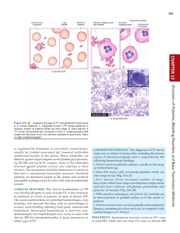
           Figure 13.6  Laboratory findings of ITP contrasted with those found
           in a normal individual. A, Peripheral blood in ITP shows presence of
           reduced number of platelets which are often large. B, Bone marrow in
           ITP shows characteristically increased number of megakaryocytes with
           single non-lobulated nuclei and reduced cytoplasmic granularity (inbox
           on right photomicrograph).


           is explained by formation of  anti-platelet autoantibodies,  LABORATORY FINDINGS. The diagnosis of ITP can be  Disorders of Platelets, Bleeding Disorders and Basic Transfusion Medicine
           usually by platelet-associated IgG humoral antibodies  suspected on clinical features after excluding the known
           synthesised mainly in the spleen. These antibodies are  causes of thrombocytopenia and is supported by the
           directed against target antigens on the platelet glycoproteins,  following haematologic findings:
           Gp IIb-IIIa and Gp Ib-IX complex. Some of the antibodies  1. Platelet count is markedly reduced, usually in the range
           directed against platelet surface also interfere in their  of 10,000-50,000/μl.
           function. The mechanism of platelet destruction is similar to
           that seen in autoimmune haemolytic anaemias. Sensitised  2. Blood film shows only occasional platelets which are
           platelets are destroyed mainly in the spleen and rendered  often large in size (Fig. 13.6,A).
           susceptible to phagocytosis by cells of the reticuloendothelial  3. Bone marrow shows increased number of mega-
           system.                                               karyocytes which have large non-lobulated single nuclei
                                                                 and may have reduced cytoplasmic granularity and
           CLINICAL FEATURES. The clinical manifestation of ITP  presence of vacuoles (Fig. 13.6, B).
           may develop abruptly in cases of acute ITP, or the onset may  4. With sensitive techniques, anti-platelet IgG antibody can
           be insidious as occurs in majority of cases of chronic ITP.  be demonstrated on platelet surface or in the serum of
           The usual manifestations are petechial haemorrhages, easy  patients.
           bruising, and mucosal bleeding such as menorrhagia in  5. Platelet survival studies reveal markedly reduced platelet
           women, nasal bleeding, bleeding from gums, melaena and  lifespan, sometimes less than one hour, as compared with
           haematuria. Intracranial haemorrhage is, however, rare.  normal lifespan of 7-10 days.
           Splenomegaly and hepatomegaly may occur in cases with
           chronic ITP but lymphadenopathy is quite uncommon in  TREATMENT. Spontaneous recovery occurs in 90% cases
           either type of ITP.                                 of acute ITP, while only less than 10% cases of chronic ITP

