


Page 345 - Textbook of Pathology, 6th Edition
P. 345
diastolic and systolic for 5 minutes. After deflation, the 329
number of petechiae appearing in the next 5 minutes in
2
3 cm area over the cubital fossa are counted. Presence of
more than 20 petechiae is considered a positive test. The test
is positive in increased capillary fragility as well as in
thrombocytopenia.
B. Investigation of Platelets and Platelet Function
Haemostatic disorders are commonly due to abnormalities
in platelet number, morphology or function.
1. SCREENING TESTS. The screening tests carried out for
assessing platelet-related causes are as under:
i) Peripheral blood platelet count.
ii) Skin bleeding time.
iii) Examination of fresh blood film to see the morphologic CHAPTER 13
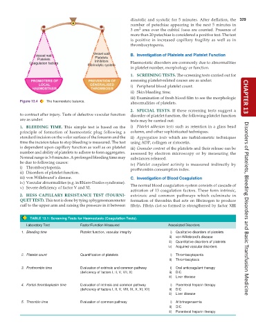
Figure 13.4 The haemostatic balance.
abnormalities of platelets.
2. SPECIAL TESTS. If these screening tests suggest a
to contract after injury. Tests of defective vascular function disorder of platelet function, the following platelet function
are as under: tests may be carried out:
1. BLEEDING TIME. This simple test is based on the i) Platelet adhesion tests such as retention in a glass bead
principle of formation of haemostatic plug following a column, and other sophisticated techniques.
standard incision on the volar surface of the forearm and the ii) Aggregation tests which are turbidometric techniques
time the incision takes to stop bleeding is measured. The test using ADP, collagen or ristocetin.
is dependent upon capillary function as well as on platelet iii) Granular content of the platelets and their release can be
number and ability of platelets to adhere to form aggregates. assessed by electron microscopy or by measuring the
Normal range is 3-8 minutes. A prolonged bleeding time may substances released.
be due to following causes: iv) Platelet coagulant activity is measured indirectly by
i) Thrombocytopenia. prothrombin consumption index.
ii) Disorders of platelet function.
iii) von Willebrand’s disease. C. Investigation of Blood Coagulation
iv) Vascular abnormalities (e.g. in Ehlers-Danlos syndrome).
v) Severe deficiency of factor V and XI. The normal blood coagulation system consists of cascade of
activation of 13 coagulation factors. These form intrinsic,
2. HESS CAPILLARY RESISTANCE TEST (TOURNI- extrinsic and common pathways which culminate in
QUET TEST). This test is done by tying sphygmomanometer formation of thrombin that acts on fibrinogen to produce
cuff to the upper arm and raising the pressure in it between fibrin. Fibrin clot so formed is strengthened by factor XIII Disorders of Platelets, Bleeding Disorders and Basic Transfusion Medicine
TABLE 13.1: Screening Tests for Haemostasis (Coagulation Tests).
Laboratory Test Factor/Function Measured Associated Disorders
1. Bleeding time Platelet function, vascular integrity i) Qualitative disorders of platelets
ii) von Willebrand’s disease
iii) Quantitative disorders of platelets
iv) Acquired vascular disorders
2. Platelet count Quantification of platelets i) Thrombocytopenia
ii) Thrombocytosis
3. Prothrombin time Evaluation of extrinsic and common pathway i) Oral anticoagulant therapy
(deficiency of factors I, II, V, VII, X) ii) DIC
iii) Liver disease
4. Partial thromboplastin time Evaluation of intrinsic and common pathway i) Parenteral heparin therapy
(deficiency of factors I, II, V, VIII, IX, X, XI, XII) ii) DIC
iii) Liver disease
5. Thrombin time Evaluation of common pathway i) Afibrinogenaemia
ii) DIC
iii) Parenteral heparin therapy

