


Page 359 - Textbook of Pathology, 6th Edition
P. 359
343


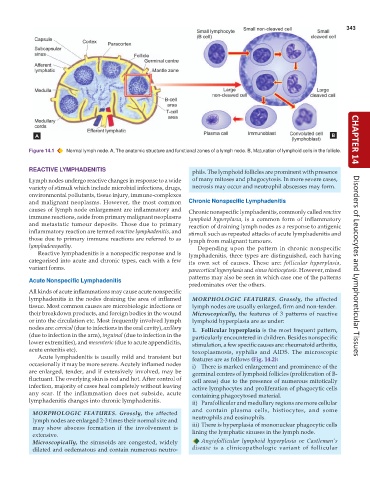
           Figure 14.1  Normal lymph node. A, The anatomic structure and functional zones of a lymph node. B, Maturation of lymphoid cells in the follicle.  CHAPTER 14


           REACTIVE LYMPHADENITIS                                phils. The lymphoid follicles are prominent with presence
           Lymph nodes undergo reactive changes in response to a wide  of many mitoses and phagocytosis. In more severe cases,
           variety of stimuli which include microbial infections, drugs,  necrosis may occur and neutrophil abscesses may form.
           environmental pollutants, tissue injury, immune-complexes
           and malignant neoplasms. However, the most common   Chronic Nonspecific Lymphadenitis
           causes of lymph node enlargement are inflammatory and  Chronic nonspecific lymphadenitis, commonly called reactive
           immune reactions, aside from primary malignant neoplasms  lymphoid hyperplasia, is a common form of inflammatory
           and metastatic tumour deposits. Those due to primary  reaction of draining lymph nodes as a response to antigenic
           inflammatory reaction are termed reactive lymphadenitis, and  stimuli such as repeated attacks of acute lymphadenitis and
           those due to primary immune reactions are referred to as  lymph from malignant tumours.
           lymphadenopathy.                                       Depending upon the pattern in chronic nonspecific
              Reactive lymphadenitis is a nonspecific response and is  lymphadenitis, three types are distinguished, each having
           categorised into acute and chronic types, each with a few  its own set of causes. These are: follicular hyperplasia,
           variant forms.                                      paracortical hyperplasia and sinus histiocytosis. However, mixed  Disorders of Leucocytes and Lymphoreticular Tissues
                                                               patterns may also be seen in which case one of the patterns
           Acute Nonspecific Lymphadenitis
                                                               predominates over the others.
           All kinds of acute inflammations may cause acute nonspecific
           lymphadenitis in the nodes draining the area of inflamed  MORPHOLOGIC FEATURES. Grossly, the affected
           tissue. Most common causes are microbiologic infections or  lymph nodes are usually enlarged, firm and non-tender.
           their breakdown products, and foreign bodies in the wound  Microscopically, the features of 3 patterns of reactive
           or into the circulation etc. Most frequently involved lymph  lymphoid hyperplasia are as under:
           nodes are: cervical (due to infections in the oral cavity), axillary  1. Follicular hyperplasia is the most frequent pattern,
           (due to infection in the arm), inguinal (due to infection in the  particularly encountered in children. Besides nonspecific
           lower extremities), and mesenteric (due to acute appendicitis,  stimulation, a few specific causes are: rheumatoid arthritis,
           acute enteritis etc).                                 toxoplasmosis, syphilis and AIDS. The microscopic
              Acute lymphadenitis is usually mild and transient but  features are as follows (Fig. 14.2):
           occasionally it may be more severe. Acutely inflamed nodes  i) There is marked enlargement and prominence of the
           are enlarged, tender, and if extensively involved, may be  germinal centres of lymphoid follicles (proliferation of B-
           fluctuant. The overlying skin is red and hot. After control of  cell areas) due to the presence of numerous mitotically
           infection, majority of cases heal completely without leaving  active lymphocytes and proliferation of phagocytic cells
           any scar. If the inflammation does not subside, acute  containing phagocytosed material.
           lymphadenitis changes into chronic lymphadenitis.     ii) Parafollicular and medullary regions are more cellular
                                                                 and contain plasma cells, histiocytes, and some
            MORPHOLOGIC FEATURES.  Grossly, the affected
            lymph nodes are enlarged 2-3 times their normal size and  neutrophils and eosinophils.
            may show abscess formation if the involvement is     iii) There is hyperplasia of mononuclear phagocytic cells
            extensive.                                           lining the lymphatic sinuses in the lymph node.
            Microscopically, the sinusoids are congested, widely    Angiofollicular lymphoid hyperplasia or  Castleman’s
            dilated and oedematous and contain numerous neutro-  disease is a clinicopathologic variant of follicular

