


Page 360 - Textbook of Pathology, 6th Edition
P. 360
344


     SECTION II
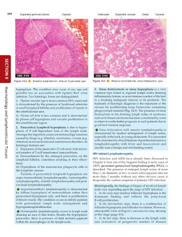
           Figure 14.2  Reactive lymphadenitis, follicular hyperplasia type.  Figure 14.3  Reactive lymphadenitis, sinus histiocytosis  type.

            hyperplasia. The condition may occur at any age and  3. Sinus histiocytosis or sinus hyperplasia is a very
            possibly has an association with Epstein-Barr virus  common type found in regional lymph nodes draining
            infection. Two histologic forms are distinguished:   inflammatory lesions, or as an immune reaction of the host
            i) Hyaline-vascular type is more common (90% cases) and  to a draining malignant tumour or its products. The
            is characterised by the presence of hyalinised arterioles  hallmark of histologic diagnosis is the expansion of the
            in small lymphoid follicles and proliferation of vessels in  sinuses by proliferating large histiocytes containing
            the interfollicular area.                            phagocytosed material (Fig. 14.3). The presence of sinus
            ii) Plasma cell form is less common and is characterised  histiocytosis in the draining lymph nodes of carcinoma
            by plasma cell hyperplasia and vascular proliferation in  such as in breast carcinoma has been considered by some
            the interfollicular region.                          workers to confer better prognosis in such patients due to
                                                                 good host immune response.
            2. Paracortical lymphoid hyperplasia is due to hyper-
            plasia of T-cell-dependent area of the lymph node.      Sinus histiocytosis with massive lymphadenopathy is
            Amongst the important causes are immunologic reactions  characterised by marked enlargement of lymph nodes,
            caused by drugs (e.g. dilantin), vaccination, viruses (e.g.  especially of the neck, in young adolescents. It is associated
            infectious mononucleosis) and autoimmune disorders. Its  with characteristic clinical features of painless but massive
            histologic features are:                             lymphadenopathy with fever and leucocytosis and
            i) Expansion of the paracortex (T-cell area) with increa-  usually runs a benign and self-limiting course.
            sed number of T-cell transformed immunoblasts.     HIV-related Lymphadenopathy
            ii) Encroachment by the enlarged paracortex on the
            lymphoid follicles, sometimes resulting in their efface-  HIV infection and AIDS have already been discussed in
     Haematology and Lymphoreticular Tissues
            ment.                                              Chapter 4; here one of the frequent finding in early cases of
            iii) Hyperplasia of the mononuclear phagocytic cells in  AIDS, persistent generalised lymphadenopathy (PGL), is
            the lymphatic sinuses.                             described. The presence of enlarged lymph nodes of more
               Variants of paracortical lymphoid hyperplasia are  than 1 cm diameter at two or more extra-inguinal sites for
            angio-immunoblastic lymphadenopathy, dermatopathic  more than 3 months without any other obvious cause is
            lymphadenopathy, dilantin lymphadenopathy and post-  frequently the earliest symptom of primary HIV infection.
            vaccinial lymphadenopathy.                           Histologically, the findings at biopsy of involved lymph
               Angioimmunoblastic lymphadenopathy is characterised  node vary depending upon the stage of HIV infection:
            by diffuse hyperplasia of immunoblasts rather than   1. In the early stage marked follicular hyperplasia is the
            paracortical hyperplasia only, and there is proliferation  dominant finding and reflects the polyclonal
            of blood vessels. The condition occurs in elderly patients  B-cell proliferation.
            with generalised lymph node enlargement and          2  In the intermediate stage, there is a combination of
            hypergammaglobulinaemia.                             follicular hyperplasia and follicular involution. However,
               Dermatopathic lymphadenopathy occurs in lymph node  adenopathic form of Kaposi’s sarcoma too may develop
            draining an area of skin lesion. Besides the hyperplastic  at this stage (page 414).
            paracortex, there is presence of dark melanin pigment  3. In the last stage, there is decrease in the lymph node
            within the macrophages in the lymph node.            size indicative of prognostic marker of disease

