


Page 434 - Textbook of Pathology, 6th Edition
P. 434
418 fibres transmit impulses from the AV node to the ventricular  adjacent half of the posterior wall of the left ventricle and
           walls.                                              the posterior third of the interventricular septum.
              The  pericardium consists of a closely apposed layer,  There are 3 anatomic patterns of distribution of the
           visceral pericardium or epicardium, and an outer fibrous sac,  coronary blood supply, depending upon which of the
           the  parietal pericardium. The two layers enclose a narrow  coronary arteries crosses the crux. Crux is the region on the
           pericardial cavity which is lined by mesothelial cells and  posterior surface of the heart where all the four cardiac
           normally contains 10-30 ml of clear, watery serous fluid. This  chambers and the interatrial and interventricular septa meet.
           fluid functions as lubricant and shock absorbant to the heart.  These patterns are as under:
              The endocardium is the smooth shiny inner lining of the  Right coronary artery preponderance is the most
           myocardium that covers all the cardiac chambers, the cardiac  common pattern. In this, right coronary artery supplies blood
           valves, the chordae tendineae and the papillary muscles. It  to the whole of right ventricle, the posterior half of the
           is lined by endothelium with connective tissue and elastic  interventricular septum and a part of the posterior wall of
           fibres in its deeper part.                          the left ventricle by crossing the crux.
              The valve cusps and semilunar leaflets are delicate and
                                                                  Balanced cardiac circulation is the next most frequent
           translucent structures. The valves are strengthened by  pattern. In this, the right and left ventricles receive blood
           collagen and elastic tissue and covered by a layer of  supply entirely from right and left coronary arteries
           endothelium (valvular endocardium).
                                                               respectively. The posterior part of the interventricular septum
           MYOCARDIAL BLOOD SUPPLY. The cardiac muscle, in     is supplied by a branch of the right coronary while the
           order to function properly, must receive adequate supply of  anterior part is supplied by a branch of the left coronary
           oxygen and nutrients. Blood is transported to myocardial  artery.
           cells by the coronary arteries which originate immediately  Left coronary preponderance is the least frequent pattern.
           above the aortic semilunar valve. Most of blood flow to the  In this, the left coronary artery supplies blood to the entire
           myocardium occurs during diastole. There are three major  left ventricle, whole of interventricular septum and also
           coronary trunks, each supplying blood to specific segments  supplies blood to a part of the posterior wall of the right
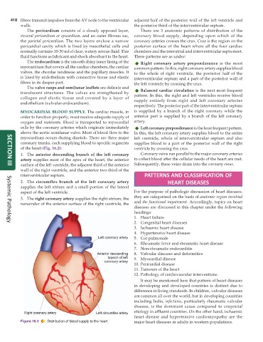
           of the heart (Fig. 16.2):                           ventricle by crossing the crux.
           1. The anterior descending branch of the left coronary  Coronary veins run parallel to the major coronary arteries
           artery supplies most of the apex of the heart, the anterior  to collect blood after the cellular needs of the heart are met.
           surface of the left ventricle, the adjacent third of the anterior  Subsequently, these veins drain into the coronary sinus.
     SECTION III
           wall of the right ventricle, and the anterior two-third of the
           interventricular septum.                                 PATTERNS AND CLASSIFICATION OF
           2. The  circumflex branch of the left coronary artery               HEART DISEASES
           supplies the left atrium and a small portion of the lateral
           aspect of the left ventricle.                       For the purpose of pathologic discussion of heart diseases,
           3. The right coronary artery supplies the right atrium, the  they are categorised on the basis of anatomic region involved
           remainder of the anterior surface of the right ventricle, the  and the functional impairment. Accordingly, topics on heart
                                                               diseases are discussed in this chapter under the following
                                                               headings:
                                                               1. Heart failure
                                                               2. Congenital heart diseases
     Systemic Pathology
                                                               3. Ischaemic heart disease
                                                               4. Hypertensive heart disease
                                                               5. Cor pulmonale
                                                               6. Rheumatic fever and rheumatic heart disease
                                                               7. Non-rheumatic endocarditis
                                                               8. Valvular diseases and deformities
                                                               9. Myocardial disease
                                                               10. Pericardial disease
                                                               11. Tumours of the heart
                                                               12. Pathology of cardiovascular interventions.
                                                                  It may be mentioned here that pattern of heart diseases
                                                               in developing and developed countries is distinct due to
                                                               difference in living standards. In children, valvular diseases
                                                               are common all over the world, but in developing countries
                                                               including India, infections, particularly rheumatic valvular
                                                               disease, is the dominant cause compared to  congenital
                                                               etiology in affluent countries. On the other hand, ischaemic
                                                               heart disease and hypertensive cardiomyopathy are the
           Figure 16.2  Distribution of blood supply to the heart.  major heart diseases in adults in western populations.

