


Page 485 - Textbook of Pathology, 6th Edition
P. 485
469


                                                                                                                      The Respiratory System


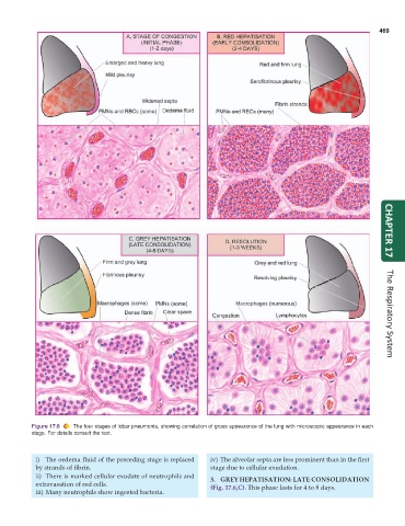
           Figure 17.6  The four stages of lobar pneumonia, showing correlation of gross appearance of the lung with microscopic appearance in each  CHAPTER 17
           stage. For details consult the text.


            i) The oedema fluid of the preceding stage is replaced  iv) The alveolar septa are less prominent than in the first
            by strands of fibrin.                                stage due to cellular exudation.
            ii) There is marked cellular exudate of neutrophils and  3. GREY HEPATISATION: LATE CONSOLIDATION
            extravasation of red cells.                          (Fig. 17.6,C). This phase lasts for 4 to 8 days.
            iii) Many neutrophils show ingested bacteria.

