


Page 490 - Textbook of Pathology, 6th Edition
P. 490
474 characteristic neutrophilic leucocytosis. Chest radiograph Legionella Pneumonia
may show patchy or diffuse consolidation. Cold agglutinin Legionella pneumonia or Legionnaire’s disease is an epidemic
titres in the serum are elevated in almost half the cases of
mycoplasmal pneumonia, 20% cases of adenovirus infection illness caused by gram-negative bacilli, Legionella pneumophila
that thrives in aquatic environment. It was first recognised
but absent in other forms of viral pneumonia. Isolation of following investigation into high mortality among those
the etiologic agent, otherwise, is difficult.
attending American Legion Convention in Philadelphia in
July 1976. The epidemic occurs in summer months by spread
C. OTHER TYPES OF PNEUMONIAS
of organisms through contaminated drinking water or in air-
Some other types of pneumonias caused by infective agents conditioning cooling towers. Impaired host defenses in the
(such as Pneumocystis carinii pneumonia and Legionella form of immunodeficiency, corticosteroid therapy, old age
pneumonia) and certain non-infective varieties (e.g. aspiration and cigarette smoking play important roles.
pneumonia, hypostatic pneumonia and lipid pneumonia) are
described here. MORPHOLOGIC FEATURES. Grossly, there are chan-
ges of widespread bronchopneumonia involving many
Pneumocystis carinii Pneumonia lobes and there may be consolidation of the entire lung.
Pneumocystis carinii, a protozoon widespread in the Pleural effusion is frequently present.
environment, causes pneumonia by inhalation of the Histologically, the changes are not distinctive. Common
organisms as an opportunistic infection in neonates and features are as under:
immunosuppressed people. Almost 100% cases of HIV/ i) Intra-alveolar exudate, initially of neutrophils but later
AIDS develop opportunistic infection during the course of composed mainly of macrophages.
disease, most commonly Pneumocystis carinii pneumonia. ii) Alveolar septa show foci of hyperplasia of the lining
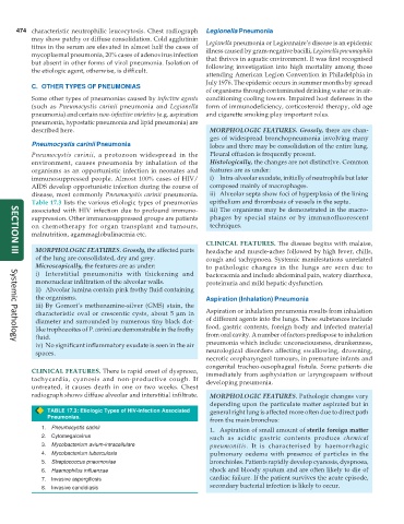
Table 17.3 lists the various etiologic types of pneumonias epithelium and thrombosis of vessels in the septa.
associated with HIV infection due to profound immuno- iii) The organisms may be demonstrated in the macro-
suppression. Other immunosuppressed groups are patients phages by special stains or by immunofluorescent
on chemotherapy for organ transplant and tumours, techniques.
malnutrition, agammaglobulinaemia etc.
CLINICAL FEATURES. The disease begins with malaise,
MORPHOLOGIC FEATURES. Grossly, the affected parts headache and muscle-aches followed by high fever, chills,
of the lung are consolidated, dry and grey. cough and tachypnoea. Systemic manifestations unrelated
SECTION III
Microscopically, the features are as under: to pathologic changes in the lungs are seen due to
i) Interstitial pneumonitis with thickening and bacteraemia and include abdominal pain, watery diarrhoea,
mononuclear infiltration of the alveolar walls. proteinuria and mild hepatic dysfunction.
ii) Alveolar lumina contain pink frothy fluid containing
the organisms. Aspiration (Inhalation) Pneumonia
iii) By Gomori’s methenamine-silver (GMS) stain, the
characteristic oval or crescentic cysts, about 5 μm in Aspiration or inhalation pneumonia results from inhalation
diameter and surrounded by numerous tiny black dot- of different agents into the lungs. These substances include
like trophozoites of P. carinii are demonstrable in the frothy food, gastric contents, foreign body and infected material
fluid. from oral cavity. A number of factors predispose to inhalation
Systemic Pathology
iv) No significant inflammatory exudate is seen in the air pneumonia which include: unconsciousness, drunkenness,
spaces. neurological disorders affecting swallowing, drowning,
necrotic oropharyngeal tumours, in premature infants and
congenital tracheo-oesophageal fistula. Some patients die
CLINICAL FEATURES. There is rapid onset of dyspnoea, immediately from asphyxiation or laryngospasm without
tachycardia, cyanosis and non-productive cough. If developing pneumonia.
untreated, it causes death in one or two weeks. Chest
radiograph shows diffuse alveolar and interstitial infiltrate. MORPHOLOGIC FEATURES. Pathologic changes vary
depending upon the particulate matter aspirated but in
TABLE 17.3: Etiologic Types of HIV-Infection Associated general right lung is affected more often due to direct path
Pneumonias.
from the main bronchus:
1. Pneumocystis carinii 1. Aspiration of small amount of sterile foreign matter
2. Cytomegalovirus such as acidic gastric contents produce chemical
3. Mycobacterium avium-intracellulare pneumonitis. It is characterised by haemorrhagic
4. Mycobacterium tuberculosis pulmonary oedema with presence of particles in the
5. Streptococcus pneumoniae bronchioles. Patients rapidly develop cyanosis, dyspnoea,
6. Haemophilus influenzae shock and bloody sputum and are often likely to die of
7. Invasive aspergillosis cardiac failure. If the patient survives the acute episode,
8. Invasive candidiasis secondary bacterial infection is likely to occur.

