


Page 487 - Textbook of Pathology, 6th Edition
P. 487
471


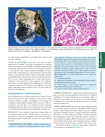
           Figure 17.9  Lobar pneumonia, grey hepatisation stage. A, The sectioned surface of the lung shows grey-brown, firm area of consolidation
           (liver-like) affecting a lobe (arrow). B, The cellular exudates in the alveolar lumina is lying separated from the septal walls by a clear space. The
           infiltrate in the lumina is composed of neutrophils and macrophages.


           are otitis media, mastoiditis, meningitis, brain abscess and  consolidation affecting one or more lobes, frequently
           purulent arthritis.                                   found bilaterally and more often involving the lower zones  CHAPTER 17
           CLINICAL FEATURES.  Classically, the onset of lobar   of the lungs due to gravitation of the secretions. On cut
           pneumonia is sudden. The major symptoms are: shaking  surface, these patchy consolidated lesions are dry,
           chills, fever, malaise with pleuritic chest pain, dyspnoea and  granular, firm, red or grey in colour, 3 to 4 cm in diameter,
           cough with expectoration which may be mucoid, purulent  slightly elevated over the surface and are often centred
           or even bloody. The common physical findings are fever,  around a bronchiole (Fig. 17.10). These patchy areas are
           tachycardia, and tachypnoea, and sometimes cyanosis if the  best picked up by passing the fingertips on the cut surface.
           patient is severely hypoxaemic. There is generally a marked  Histologically, the following features are observed
           neutrophilic leucocytosis. Blood cultures are positive in about  (Fig. 17.11):
           30% of cases. Chest radiograph may reveal consolidation.  i) Acute bronchiolitis.
           Culture of the organisms in the sputum and antibiotic  ii) Suppurative exudate, consisting chiefly of neutrophils,  The Respiratory System
           sensitivity are most significant investigations for institution  in the peribronchiolar alveoli.
           of specific antibiotics. The response to antibiotics is usually  iii) Thickening of the alveolar septa by congested
           rapid with clinical improvement in 48 to 72 hours after the  capillaries and leucocytic infiltration.
           initiation of antibiotics.                            iv) Less involved alveoli contain oedema fluid.

           Bronchopneumonia  (Lobular Pneumonia)               COMPLICATIONS. The complications of lobar pneumonia
                                                               may occur in bronchopneumonia as well. However, complete
           Bronchopneumonia or lobular pneumonia is infection of the  resolution of bronchopneumonia is uncommon. There is
           terminal bronchioles that extends into the surrounding  generally some degree of destruction of the bronchioles
           alveoli resulting in patchy consolidation of the lung. The  resulting in foci of bronchiolar fibrosis that may eventually
           condition is particularly frequent at the extremes of life (i.e.  cause bronchiectasis.
           in infancy and old age), as a terminal event in chronic
           debilitating diseases and as a secondary infection following  CLINICAL FEATURES. The patients of bronchopneumonia
           viral respiratory infections such as influenza, measles etc.  are generally infants or elderly individuals. There may be
                                                               history of preceding bed-ridden illness, chronic debility,
           ETIOLOGY.  The common organisms responsible for     aspiration of gastric contents or upper respiratory infection.
           bronchopneumonia are staphylococci, streptococci,   For initial 2 to 3 days, there are features of acute bronchitis
           pneumococci, Klebsiella pneumoniae, Haemophilus influenzae,  but subsequently signs and symptoms similar to those of
           and gram-negative bacilli like  Pseudomonas and coliform  lobar pneumonia appear. Blood examination usually shows
           bacteria.                                           a neutrophilic leucocytosis. Chest radiograph shows mottled,
                                                               focal opacities in both the lungs, chiefly in the lower zones.
            MORPHOLOGIC FEATURES. Grossly, bronchopneu-           The salient features of the two main types of bacterial
            monia is identified by patchy areas of red or grey
                                                               pneumonias are contrasted in Table 17.2.

